39 sarcomere structure labeled
Diagram Of Sarcomere Diagram Of Sarcomere A sarcomere is the basic unit of striated muscle tissue. It is the repeating unit between two Z lines. Skeletal muscles are composed of tubular muscle cells which. sarcomere. Schematic: The Z line is depicted in black, myosin in red, actin in green/gray, and tropomyosin in blue. Image: MPI of Molecular Plant Physiology. Sarcomere - an overview | ScienceDirect Topics A sarcomere is the basic contractile unit of muscle fiber. Each sarcomere is composed of two main protein filaments—actin and myosin—which are the active structures responsible for muscular contraction. The most popular model that describes muscular contraction is called the sliding filament theory.
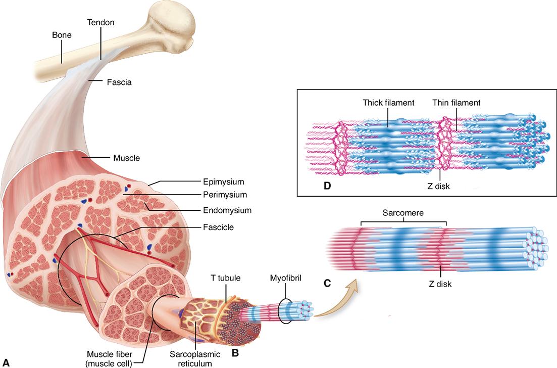
10.2 Skeletal Muscle - Anatomy & Physiology A sarcomere is defined as the region of a myofibril contained between two cytoskeletal structures called Z-discs (also called Z-lines), and the striated appearance of skeletal muscle fibers is due to the arrangement of the thick and thin myofilaments within each sarcomere ( Figure 10.2.2 ).

Sarcomere structure labeled
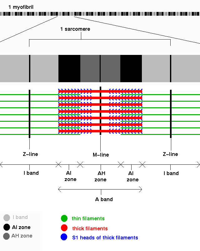
Sarcomere Diagram Labeled - schematron.org Sarcomeres are composed of thick filaments and thin filaments. The thin filaments Look at the diagram above and realize what happens as a muscle contracts. Draw your own diagram of two sarcomeres. The first should be of a relaxed muscle. The second should be of a contracted muscle. Label the Z line, M line. Sarcomere: Structure and Parts, Functions and Histology The main components of the histology of a sarcomere are summarized below: Band A Thick filament zone, composed of myosin proteins. Zone H Central zone of band A, without actin proteins superimposed when the muscle is relaxed. Band I Zone of thin filaments, composed of actin proteins (without myosin). Z disks Labeled Sarcomere Diagram - schematron.org Sarcomeres are composed of thick filaments and thin filaments. The thin filaments Look at the diagram above and realize what happens as a muscle contracts. Each myofibril is made up of contractile sarcomeres AND Drawing labelled diagrams of the structure of a sarcomere. A sarcomere is the basic unit of striated muscle tissue.
Sarcomere structure labeled. Sarcomere Model Sarcomere Structure - YouTube This video was produced to help students of human anatomy at Modesto Junior College study our anatomical models. Sarcomere Labeling Diagram | Quizlet Sarcomere The smallest contractile unit of muscle; extends from one Z disc to the next H Band The band at the middle of the A Band, where only myosin is found A Band The darkest area that runs the length of the myosin, including where actin and myosin overlap I Band On either side of the A Band is the I band, where only the Actin is found Z Disk Label the Sarcomere Structure Diagram | Quizlet Only $35.99/year Label the Sarcomere Structure STUDY Learn Write Test PLAY Match Created by jack_burton76PLUS Terms in this set (12) z disc mysosin (thick) thin (actin) filament I band A band I band H zone elastic (titin) filaments elastic (titin) filaments thin (actin) filament thick (myosin) filaments myosin heads Sets found in the same folder Sarcomere - an overview | ScienceDirect Topics A sarcomere is the basic contractile unit of muscle fiber. Each sarcomere is composed of two main protein filaments—actin and myosin—which are the active structures responsible for muscular contraction. The most popular model that describes muscular contraction is called the sliding filament theory.
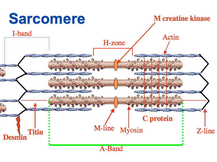
Sarcomere Structure : Mnemonic | Epomedicine Z is the final alphabet: Z lines represents the end of sarcomere M for middle: M line represents the midline of sarcomere I is a thin letter: I band has only thin filaments H is a thick letter: H zone has only thick filaments A is a hybrid of "I" and "H": A band has both thin and thick filaments (remains constant during contraction) Sarcomere (Muscle) Coloring - The Biology Corner The enter muscle fiber is surrounded by the sarcolemma (D), color this membrane brown. If expanded, the light and dark bands are shown as individual thick and thin filaments. Color the thick filaments (not labeled) red and the thin filaments blue. The Z line is the boundary between sarcomeres, named after its shape. Color the Z-line orange. Sarcomere: Structure and Parts, Functions and Histology Each sarcomere consists of thick and thin bundles of the proteins mentioned above, which together are called myofilaments. By enlarging a portion of the myofilaments, the molecules that compose them can be identified. The thick filaments are made of myosin, while the fine filaments are made of actin. Sarcomere: anatomy, structure and function | Kenhub The structure of the sarcomere is traditionally described with dark and light bands visible under the microscope. This banding pattern in sarcomeres is due mainly to the arrangement of thick and thin myofilaments in each unit. These markings include: A bands (or anisotropic bands) - dark bands that contain whole thick filaments (myosin).
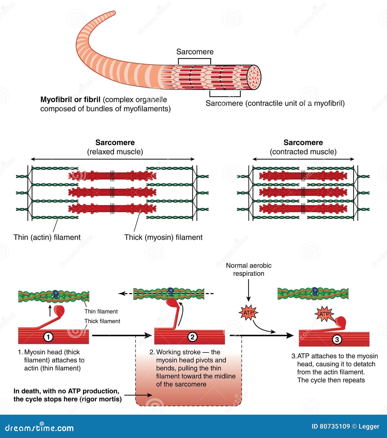
Contracted Sarcomere Diagram Draw your own diagram of two sarcomeres. The first should be of a relaxed muscle. The second should be of a contracted muscle. Label the Z line, M line. (B) A conceptual diagram representing the connectivity of molecules within a sarcomere. A person Comparison of a relaxed and contracted sarcomere. The contraction of a striated muscle fiber ... Sarcomere - Definition, Structure, Function and Quiz - Biology Dictionary Sarcomeres are able to initiate large, sweeping movement by contracting in unison. Their unique structure allows these tiny units to coordinate our muscles' contractions. The image depicts skeletal muscle fiber. In fact, the contractile properties of muscle are a defining characteristic of animals. Animal movement is notably smooth and complex. Sarcomere - Muscle Contraction - SmartDraw Skeletal muscle, also called striated muscle tissue, is made up of a series of sarcomeres. A sarcomere consists of myosin and actin filaments which overlap upon contraction. Myosin bonds with actin to ratchet the tropomyosin down the length of the myosin. Sarcomere. Actin filament. Labeled Sarcomere Diagram A sarcomere is the basic unit of striated muscle tissue. It is the repeating unit between two Z lines. Skeletal muscles are composed of tubular muscle cells which. Sarcomeres are composed of thick filaments and thin filaments. The thin filaments Look at the diagram above and realize what happens as a muscle contracts.
Physiology of the Muscular System | Basicmedical Key
Anatomy of the cardiac sarcomere. (A) Diagram of the basic organization ... The sarcomere forms the basic contractile unit in the cardiomyocytes of the heart. Thin filaments composed of actin are anchored at the Z line and form transient sliding interactions with thick ...
PPS '96: Muscle Fibres Part 2
Describe the structure of sarcomere. - Toppr Ask Sarcomeres are composed of long, fibrous proteins as filaments that slide past each other when a muscle contracts or relaxes. 1) Myosin forms the thick filament. Myosin has a long fibrous tail and a globular head, which binds to actin. Its head also binds to ATP, which is the source of energy for muscle movement. 2) Actin forms the thin filament.
Contraction Cartoons, Illustrations & Vector Stock Images - 2823 ...
Sliding Filament - TeachMeAnatomy - Making Anatomy Simple Ultrastructural Appearance of Skeletal Muscle. The striated appearance of skeletal muscle fibres is due to the organisation of two contractile proteins: actin (thin filament) and myosin (thick filament). The functional unit of contraction in a skeletal muscle fibre is the sarcomere, which runs from Z line to Z line.

Skeletal Muscle Histology - Embryology
Sarcomere Diagram Labeled - Wiring Diagrams As will soon be described, the functional unit of a skeletal muscle fiber is the sarcomere, a highly organized arrangement of the contractile myofilaments actin . Draw your own diagram of two sarcomeres. The first should be of a relaxed muscle. The second should be of a contracted muscle. Label the Z line, M line.

Sarcomere
Labeled Sarcomere Diagram - schematron.org Sarcomeres are composed of thick filaments and thin filaments. The thin filaments Look at the diagram above and realize what happens as a muscle contracts. Each myofibril is made up of contractile sarcomeres AND Drawing labelled diagrams of the structure of a sarcomere. A sarcomere is the basic unit of striated muscle tissue.
Gross Muscle and Sarcomere
Sarcomere: Structure and Parts, Functions and Histology The main components of the histology of a sarcomere are summarized below: Band A Thick filament zone, composed of myosin proteins. Zone H Central zone of band A, without actin proteins superimposed when the muscle is relaxed. Band I Zone of thin filaments, composed of actin proteins (without myosin). Z disks

Skeletal muscle fiber model
Sarcomere Diagram Labeled - schematron.org Sarcomeres are composed of thick filaments and thin filaments. The thin filaments Look at the diagram above and realize what happens as a muscle contracts. Draw your own diagram of two sarcomeres. The first should be of a relaxed muscle. The second should be of a contracted muscle. Label the Z line, M line.

Complete Giant Sarcomere Model by Denoyer-Geppert - Anatomy Models and ...

The skeletal muscle fiber stock vector. Illustration of normal - 41221577
Post a Comment for "39 sarcomere structure labeled"