41 labelled electron micrograph of chloroplast
Plant Cell Nucleus Electron Micrograph : Cell And Organelles Dr Jastrow ... The nucleus (plural = nuclei) figure 7.14 at left a transmission electron micrograph and at right a labeled diagram of a. An electron micrograph of a cell nucleus showing a densely staining nucleolus. Plant cell, electron micrograph 13 plant cells and tissues 29, 30 fiber 11. Plant cells also contain chloroplasts, often have a large, permanent ... Electron Micrographs** Electron Micrographs**. Below is a collection of electron micrographs with labelled subcellular structures that you should be able to identify. Also, be sure to observe any electron micrographs which are made available in the laboratory by the instructor. You should concentrate on the similarities in form that permit identification of the ...
Draw a labelled diagram of chloroplast as seen under an electron ... Click here👆to get an answer to your question ️ Draw a labelled diagram of chloroplast as seen under an electron microscope. Name the three major photosynthetic pigments. Solve Study Textbooks Guides. ... Draw a labelled diagram of chloroplast as seen under an electron microscope. Name the three major photosynthetic pigments. Hard. Open in App.
Labelled electron micrograph of chloroplast
PDF Question paper (AS) : Paper 1 - June 2019 - AQA Figure 3 is an electron micrograph of a chloroplast. Figure 3 0 5 . 2 Identify structures labelled [2 marks] ... Figure 4is an image of a fish gill taken using a scanning electron microscope. Figure 4 . 0 6 . 1 Identify structures labelled F and G. [1 mark] F G. 0 6 . 2 Label This Transmission Electron Micrograph : TEM of chloroplast from ... Provide the labels for the electron micrograph in figure 12.8. Label the transmission electron micrograph of the nucleus. Label the transmission electron micrograph of the nucleus. Transmission electron microscopy (tem) is a microscopy technique in which a beam of electrons is transmitted through a specimen to form an image. Animal Cell Electron Microscope Labelled - Q14 Draw a large diagram of ... Electron microscopes use accelerated electron beams (as opposed to visible light in a light microscope) to create images of magnification as here is an electron micrograph of an animal cell with the labels superimposed: (i) name the parts labelled as 1 to 10.
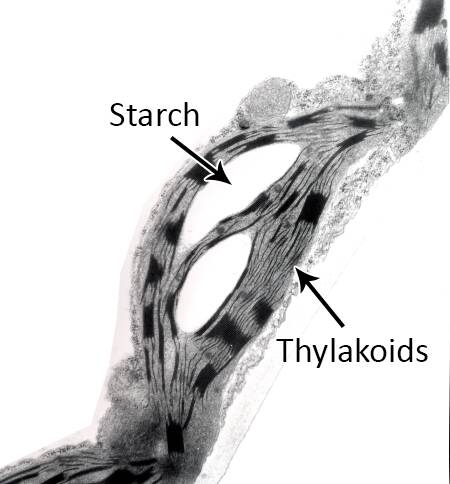
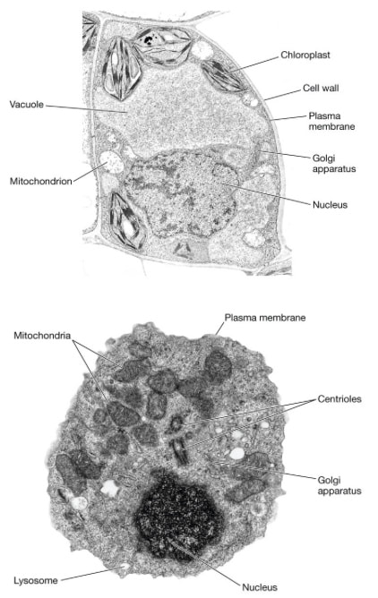
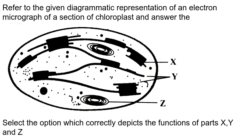
Labelled electron micrograph of chloroplast. Refer to the given diagrammatic representation of an electron ... Refer to the given diagrammatic representation of an electron micrograph of a section of chloroplast and answer the ... Refer to the given diagrammatic representation of an electron micrograph of a section of chloroplast and answer the Select the option that correct ide. asked Feb 18 in Biology by AkashBansal (37.3k points) class-11 ... PDF Identifying Organelles from an Electron Micrograph Courtesy of Dr. Julian Thorpe - EM & FACS Lab, Biological Sciences University Of Sussex The electron micrograph displayed below illustrates many of the plant cell characteristics discussed The cell wall, large central vacuole and chloroplasts are clearly visible Also visible is the clearly defined nucleus containing chromatin Cell Biology, Chloroplast - University of Florida They are specially designed to absorb light and convert it to chemical energy. Electron micrograph of a chloroplast Chloroplasts are organelles bounded by an outer membrane, but they also have an important inner membrane system. The inner membrane system is where photosynthesis takes place. Electron micrograph of a chloroplast Chloroplast Micrograph Stock Photos and Images - Alamy Sketch of an electron micrograph of a chloroplast from Zea mais. The dark regions are the grana. They are cylinders about 4,000 to 6,000 A in diameter and 5,000 to 8,000 A in height. After E. I. Rabinowitch, Photosynthesis, II, 2 (New York: Interscience Publishers, Inc., 1956), from Vatter, unpublished, modified. and mo ID: RHJXJ3 (RM)
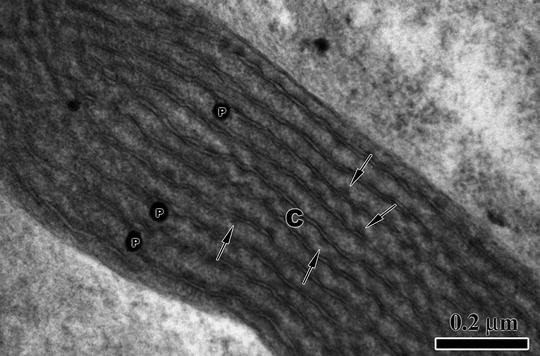
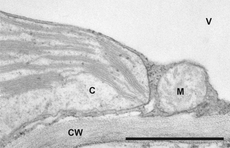
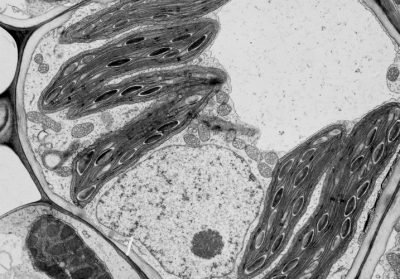
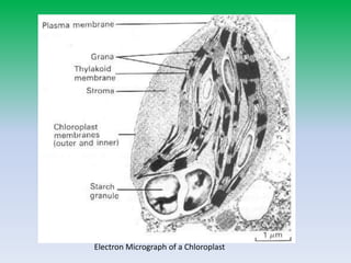
chloroplast and mitochondrion.pdf - D SBA#: Title: Electron micrograph ... SBA# Electron micrograph of a chloroplast Skilled Assessed: D (Drawing) Criteria Total Marks Marks Obtained No evidence of shading 1 Drawing is a good representation of specimen 1 Parts of drawings are proportional to parts of specimen (-1 for deviation) 2 Drawing is large enough to observe necessary details (occupies at least 50% of page) 1 ... Chloroplast- Diagram, Structure and Function Of Chloroplast It is oval or biconvex, found within the mesophyll of the plant cell. The size of the chloroplast usually varies between 4-6 µm in diameter and 1-3 µm in thickness. They are double-membrane organelle with the presence of outer, inner and intermembrane space. There are two distinct regions present inside a chloroplast known as the grana and stroma. Draw a labelled diagram of chloroplast as seen under an electron ... Draw a labelled diagram of chloroplast as seen under an electron microscope. Name the three major photosynthetic pigments. Hard Solution Verified by Toppr Chlorophyll a: Light to medium green. Main photosynthetic pigment. Chlorophyll b: Blue-green. Accessory Pigment. Carotene: Orange. Accessory Pigment. Xanthophyll: Yellow. Accessory Pigment. Transmission electron microscopic images of chloroplasts and ... For easy organelle identification, a chloroplast (P) and a mitochondrion (M) are labeled. (C-E) Ultrastructure of chloroplasts and mitochondria in cells of the strong PRORP1 RNAi mutant line RNAi-2.
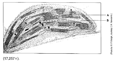
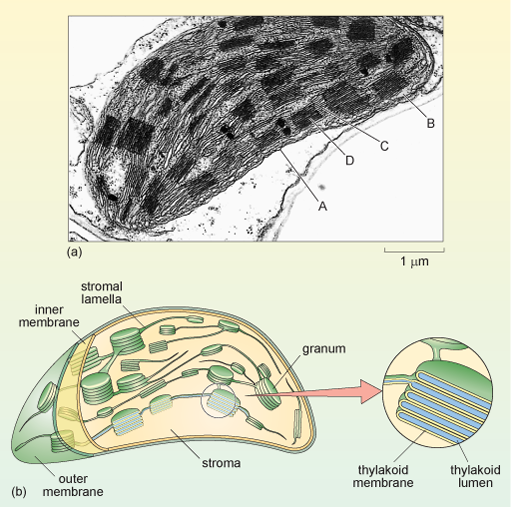
Scanning electron microscopy of chloroplast ultrastructure Abstract. A range of fracturing and sectioning techniques are now available which permit intracellular structures to be observed in the scanning electron microscope. One such technique, based on the method of Tanaka (1981), has been used to study chloroplast ultrastructure in Japan laurel, Aucuba japonica. Small pieces of leaves were fixed ... PDF Chloroplasts Structure and Function Factsheet The biconvex shape of the chloroplast is yet another way of increasing surface area to maximise absorption of light energy Sometimes in the exam you will be presented with an electron micrograph of a chloroplast. Usually, the first question simply asks you to label it. Typical Exam Question Label parts A B & C A B C Answer A - stroma; Labeling the Cell Flashcards | Quizlet Label the structures of the plasma membrane and cytoskeleton. Label the membranous organelles. ... Label the transmission electron micrograph of the mitochondrion. Label the transmission electron micrograph of the nucleus. membrane bound organelles. golgi apparatus, mitochondrion, lysosome, peroxisome, rough endoplasmic reticulum ... Chloroplasts - Biology Pages Illustrated discussion of the chloroplast genome The electron micrograph above on the right (courtesy of Dr. L. K. Shumway) shows the chloroplast from the cell of a corn leaf. The electron micrograph on the left (courtesy of Kenneth R. Miller) shows the inner surface of a thylakoid membrane. Each particle may represent one photosystem II complex.
The Structure and Morphology of Red Algae Chloroplasts ...
plant cell label electron micrograph Diagram | Quizlet Start studying plant cell label electron micrograph. Learn vocabulary, terms, and more with flashcards, games, and other study tools.

Chloroplasts (13.1.1) | CIE A Level Biology Revision Notes ...
Leaf chloroplast - Radboud Universiteit Plants utilize their leaves to produce sugars in a process called photosynthesis. Photosynthesis starts with the capture of light energy from the sun by pigments that are located in the internal membranes of chloroplasts. Chloroplasts are abundant in the sponge parenchyma of leaves. With field-emission scanning electron microscopy (FESEM) and ...
Solved Examine this electron micrograph of a chlorplast ...
IB Questionbank - examsnap.io 16M.2.HL.TZ0.7a: Draw a labelled diagram of a eukaryotic plant cell as seen in an electron micrograph. 16N.2.SL.TZ0.2a: The image is an electron micrograph. Determine, with a reason, whether the image is of a... 16N.3.SL.TZ0.3a: State from which organ the section was taken.

Warm Up 11/4/13 | Heena Bio HL YAY
Electron micrograph of isolated chloroplasts with the major organellar ... Electron micrograph of isolated chloroplasts with the major organellar subcompartments labelled. Arrows indicate immunogold-labelled preprotein that is trapped at an intermediate stage of...
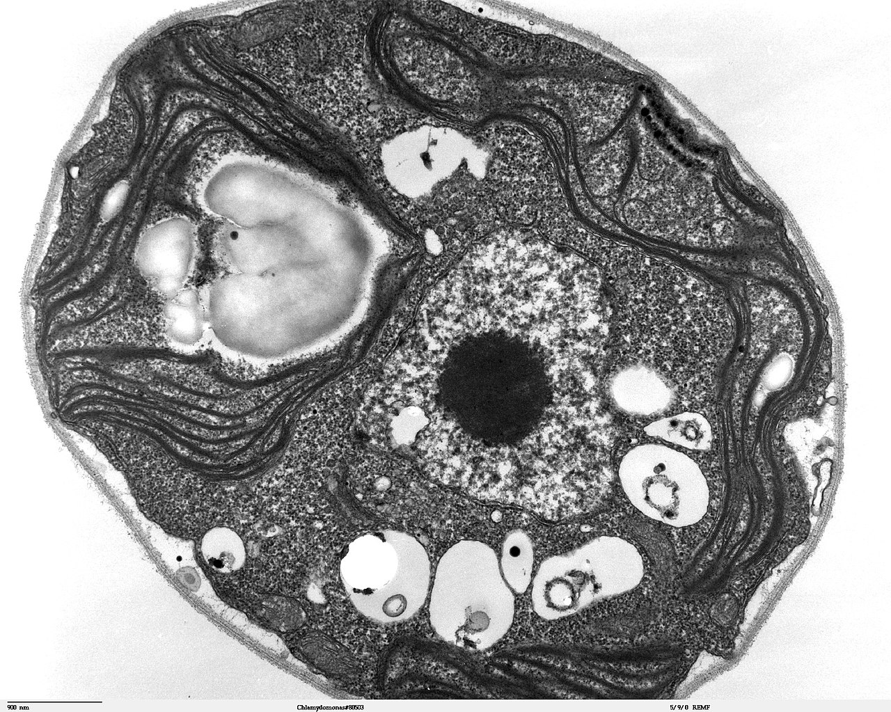
File:Chlamydomonas TEM 07.jpg - Wikimedia Commons
Solved Examine this electron micrograph of a chlorplast. - Chegg Examine this electron micrograph of a chlorplast. A. Identify the stack of membranes labeled A. B. Identify the region labeled B. C. Would the production of organic compounds during the light-independent reactions occur in; Question: Examine this electron micrograph of a chlorplast. A.

Biology: 1.2 Ultrastructure of Cells Flashcards | Quizlet
Chloroplasts - Definition, Structure, Function and Microscopy What are Chloroplasts? Essentially, chloroplasts are plastids found in cells of higher plants (plants with advanced traits with lignified tissue for transport of water and minerals) and algae as sites of photosynthesis. This makes them the most important cell organelles given that plants are the primary producers and the base of all food chains ...

Failure of the Peritoneal-Button Operation for Ascites ...
Classical transmission electron microscopy (TEM) led to the formulation ... Early electron micrographs of isolated chloroplast membranes. a Clusters of grana from individual, disrupted spinach chloroplasts air-dried on grid. The arrow points to a small, separated cluster of grana that appear interconnected by membranes (from Granick and Porter 1947). b Cluster of three, round, gold-shadowed grana "stacks" at high magnification (from Granick and Porter 1947).

Electron micrograph of chloroplasts (C) inside cells lining ...
6 examine the electron micrograph of a chloroplast a 6. Examine the electron micrograph of a chloroplast. (a) identify the stack of membranes labeled A.-Stack of thylakoids is granum (b) identify the region labeled B.-Inner aqueous fluid, stroma (c) would the production of organic compounds during the light independent reactions occur in region B or on the membranes labeled A.-Light independent reactions would only occur in region B the stroma ...
The paper
The diagram below represents a section through a chloroplast as seen ... The diagram below represents a section through a chloroplast as seen under the electron microscope. (a) Name the structure labelled D. (b) In which labelled structure do we find chlorophyll molecule. (c) Name the structure labelled, where carbon IV oxide fixation occurs.
Arabidopsis thaliana(L.) Accession Col | Science Lab | Leica ...
Electron Micrographs of Cell Organelles | Zoology (6) The main function of chloroplast or plastid is to synthesize carbohydrate molecules from CO 2 + H 2 O using light energy. 6. The Electron Micrograph of Nucleus: This is an electron micrograph of nucleus. (Fig. 17 & 18): (1) Nucleus was discovered by Brown (1831).
Detail of a chloroplast with well developed grana - UWDC - UW ...
Electron micrographs chloroplast - Big Chemical Encyclopedia The inner membrane system of the chloroplasts was found to disintegrate prior to the breakdown of other cell structures. [Pg.131] Figure 4. Electron micrograph of longitudinal sections of Chlamydomonas reinhardtii Dang. A. Control cell B. Cell treated 1 h with 1.0-1.5 g/ml nonylphenol C. Chloroplast d. dichtyosome f. flagella m. mitochondrion ...
Structure of the Chloroplast
Animal Cell Electron Microscope Labelled - Q14 Draw a large diagram of ... Electron microscopes use accelerated electron beams (as opposed to visible light in a light microscope) to create images of magnification as here is an electron micrograph of an animal cell with the labels superimposed: (i) name the parts labelled as 1 to 10.

Draw a neat labelled diagram of chloroplast.
Label This Transmission Electron Micrograph : TEM of chloroplast from ... Provide the labels for the electron micrograph in figure 12.8. Label the transmission electron micrograph of the nucleus. Label the transmission electron micrograph of the nucleus. Transmission electron microscopy (tem) is a microscopy technique in which a beam of electrons is transmitted through a specimen to form an image.
What is a diagram of a plant and animal cell under an ...
PDF Question paper (AS) : Paper 1 - June 2019 - AQA Figure 3 is an electron micrograph of a chloroplast. Figure 3 0 5 . 2 Identify structures labelled [2 marks] ... Figure 4is an image of a fish gill taken using a scanning electron microscope. Figure 4 . 0 6 . 1 Identify structures labelled F and G. [1 mark] F G. 0 6 . 2

X,Y,Z),("Dark reaction","Light reaction","Cytoplasmic ...
Relationships Between Lipid Changes and Plastid ...
Mitochondria and chloroplasts (article) | Khan Academy
3.3 Eukaryotic Cells – Concepts of Biology – 1st Canadian Edition
Chloroplast Electron Micrograph Labeled 15 Images - Quia ...

2.1 The Structure & Functions of Eukaryotic Cells - ppt download
Structure of the Chloroplast
D2: Digestion (Core) - AMAZING WORLD OF SCIENCE WITH MR. GREEN

Chloroplast Under Electron Microscope 15 Images - Structure ...
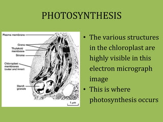
Photosynthesis

Electron micrograph of isolated chloroplasts with the major ...
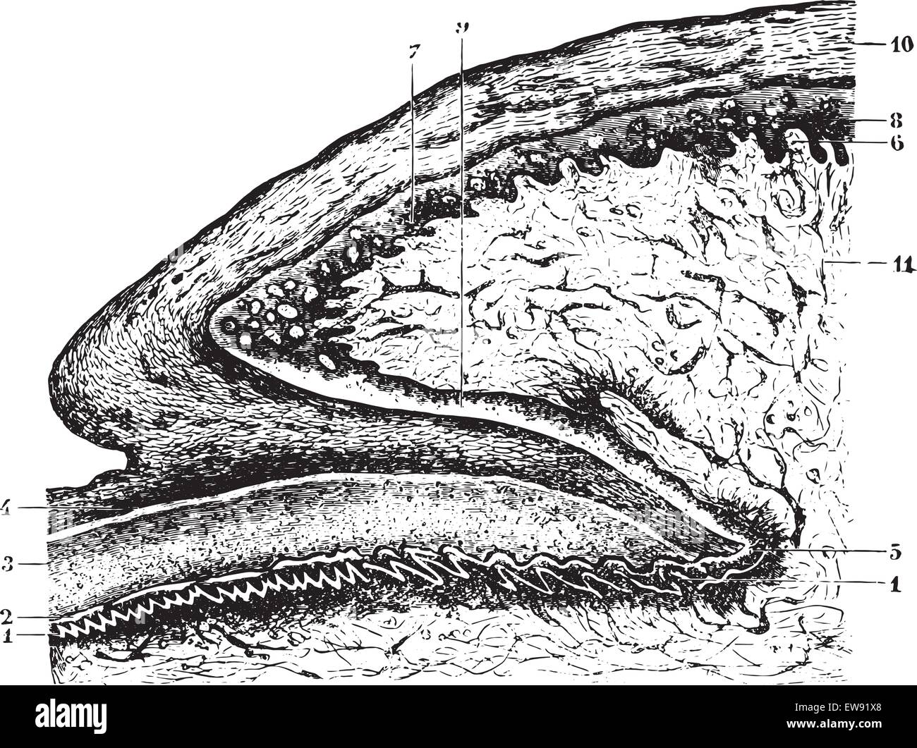
Cutting of the matrix and the root of the nail, vintage ...

Topic 1.2 Ultrastructure Of Cells - Lessons - Blendspace

1.2 Ultrastructure of Cells

Cell Micrographs | BioNinja

Electron micrograph of isolated chloroplasts with the major ...

File:Chloroplast in leaf of Anemone sp TEM 30000x.png ...
Show Question

Ultrastructure of cells 1.2

Photosynthesis Lesson 1 – review lesson for structure of a ...

DP Topic 1.1 / 1.2 | Biology - Quizizz
Sucrose or starch? - Encyclopedia of the Environment
A tour of the cell: View as single page
Photosynthesis Part 2
X,Y,Z),("Dark reaction","Light reaction","Cytoplasmic ...

Modulating the activities of chloroplasts and mitochondria ...

Cell Micrographs | BioNinja
Post a Comment for "41 labelled electron micrograph of chloroplast"