42 provide the labels for the electron micrograph
Solved Label the transmission electron micrograph based on - Chegg Solved Label the transmission electron micrograph based on | Chegg.com. Science. Biology. Biology questions and answers. Label the transmission electron micrograph based on the hints provided Mitochondrion Heterochromatin Plasma cell Nucleus Rough endoplasmic reticulum Nucleolus. PDF Identifying Organelles from an Electron Micrograph Courtesy of Dr. Julian Thorpe - EM & FACS Lab, Biological Sciences University Of Sussex The electron micrograph displayed below illustrates many of the plant cell characteristics discussed The cell wall, large central vacuole and chloroplasts are clearly visible Also visible is the clearly defined nucleus containing chromatin
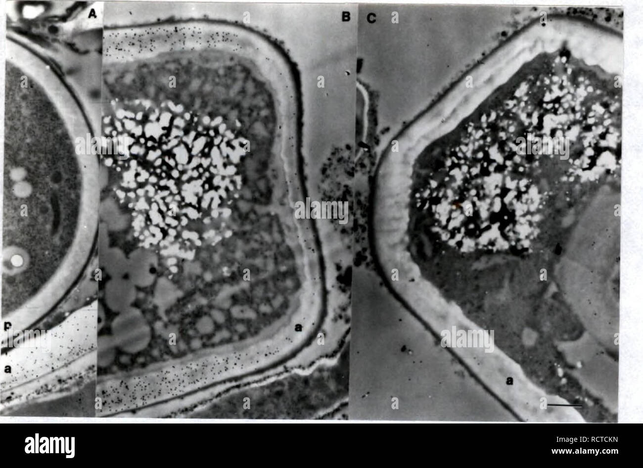
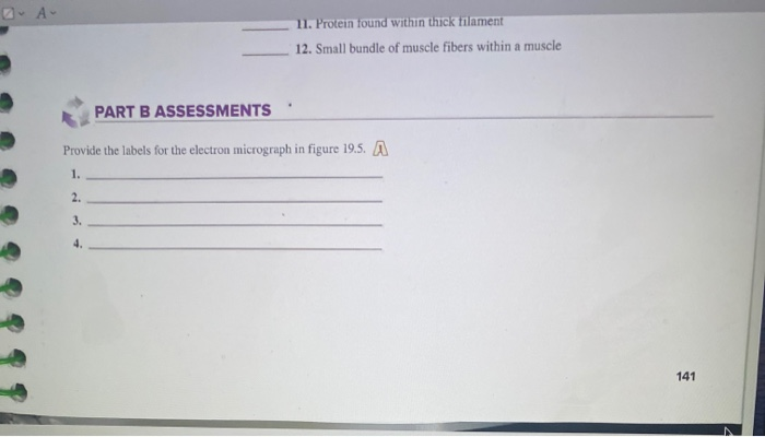
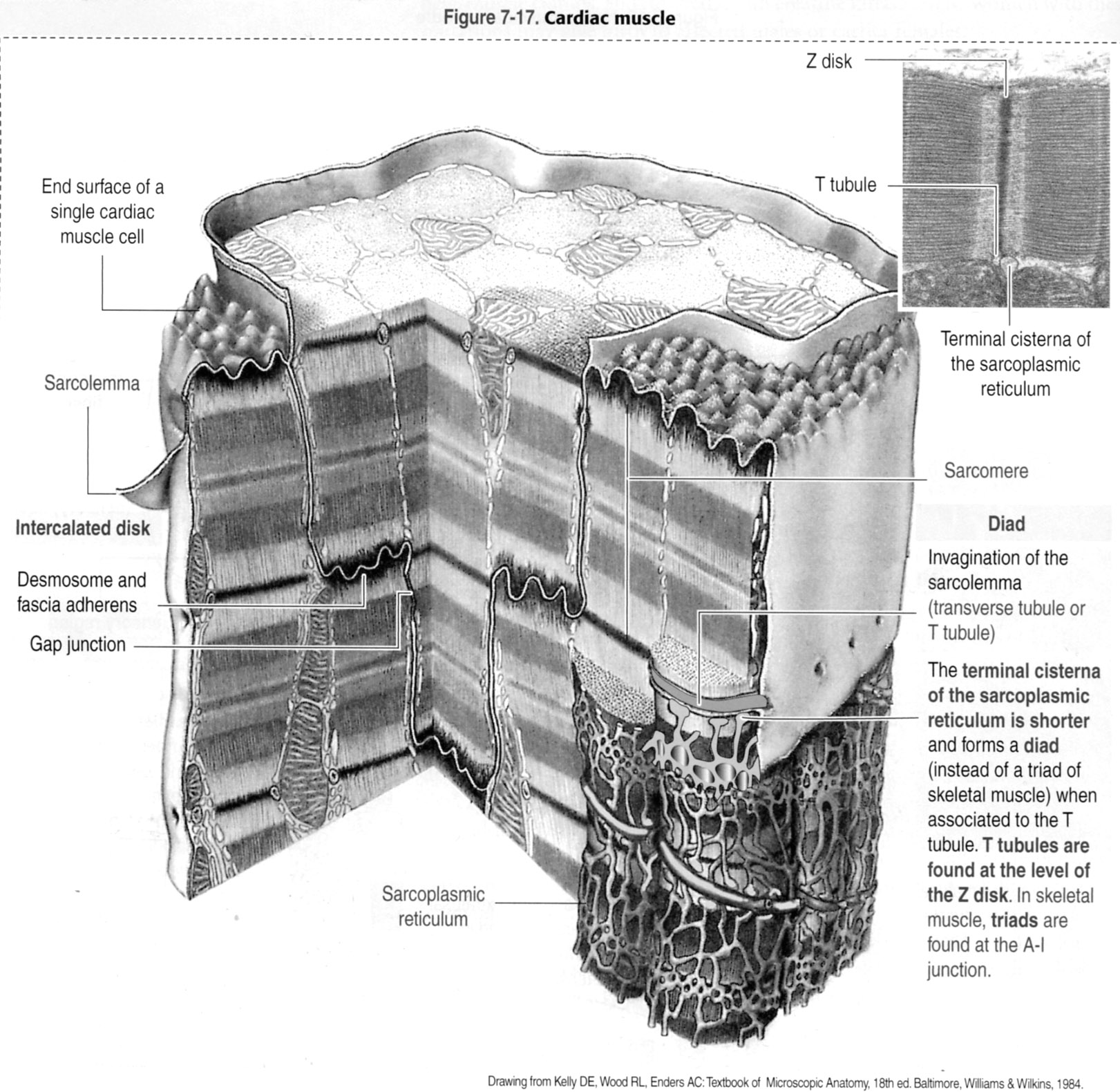
8 Human A&P | Lab Terms: A band (dark) emps I band (light ... Provide the labels for the electron micrograph in figure 19.5 Image transcription text 8 Human A&P | Lab Terms: A band (dark) emps I band (light) Sarcomere 2 Z line H zone M line 3 GURE 19.5 Using the terms provided, identify the bands and lines of the striations of a transmission electron crograph of relaxed sarcomeres (16,000x).
Provide the labels for the electron micrograph
Electron Micrographs of Cell Organelles | Zoology It is an electron micrograph of cell’s largest and most important organelle – the mitochondria and is characterized by the following features (Fig. 7 & 8): (1) The name mitochondria was given by Benda (1898) and their ma n function was brought to light by Kingsbury (1912). (2) Each mitochondria in section appears as sausage or cup or bowl ... Electron microscope - Wikipedia An electron microscope is a microscope that uses a beam of accelerated electrons as a source of illumination. As the wavelength of an electron can be up to 100,000 times shorter than that of visible light photons, electron microscopes have a higher resolving power than light microscopes and can reveal the structure of smaller objects.. Electron microscopes use shaped magnetic fields to form ... Electron Microscopy SciencesExpiration Labels | Fisher Scientific Manufacturer: Electron Microscopy Sciences 7702405. Quickly identify outdated laboratory items such as reagents and controls. These labels provide space for noting the date that the items were received, opened and when it will expire. A permanent adhesive holds the label firmly in place on a variety of surfaces. The label measures 3/4" x 1 1/2".
Provide the labels for the electron micrograph. The Apex of Protein Tags for Electron Microscopy The Apex of Protein Tags for Electron Microscopy Scientists have developed a new peroxidise-derived tag that can be used to label proteins for visualization by electron microscopy. Label the microscope — Science Learning Hub Label the microscope Add to collection Use this interactive to identify and label the main parts of a microscope. Drag and drop the text labels onto the microscope diagram. eye piece lens coarse focus adjustment high-power objective diaphragm or iris base fine focus adjustment light source stage Download Exercise Tweet What Is an Electron Microscope (EM) and How Does It Work? - VHA ... A stream of high voltage electrons (usually 5-100 KeV) is formed by the Electron Source (usually a heated tungsten or field emission filament) and accelerated in a vacuum toward the specimen using a positive electrical potential. This stream is confined and focused using metal apertures and magnetic lenses into a thin, focused, monochromatic beam. Label This Transmission Electron Micrograph Of A Relaxed … 19-03-2022 · From science 101 at university high school, tucson. Note how the sarcomeres are extended to only approximately 120 % . Provide the labels for the electron micrograph in figure 12.8. Figures label this transmission electron micrograph ( 16, 000 x ) of a relaxed sarcomere by placing the. Label This Transmission Electron Micrograph Of A Relaxed ...
Electron Micrographs** Electron Micrographs** Below is a collection of electron micrographs with labelled subcellular structures that you should be able to identify. Also, be sure to observe any electron micrographs which are made available in the laboratory by the instructor. Labels, Electron Microscopy Sciences | VWR Labels, Electron Microscopy Sciences Supplier: Electron Microscopy Sciences Labels with expanded temperature range and freezable in liquid and vapor phase nitrogen. They adhere to most plastics, glass and metals without cracking, peeling or degrading. Compatible and Economical Temperature resistant from -196 to 70 °C Color highlights detail in electron microscope images - New Atlas Color highlights detail in electron microscope images. By Michael Irving. November 08, 2016. On the left is a standard grayscale electron micrograph of a mouse brain, and on the right is a ... Multicolor Labels in Electron Microscopy [Roger Tsien ... - BioTechniques Now, however, a new paper in Cell Chemical Biology (2) reports the first steps toward a technicolor EM world: two clear, crisp, red and green labels pseudocoloring a series of elegant electron micrographs. Painting with Metals
Solved: Chapter 20 Problem 1LAB Solution - Chegg Access Laboratory Manual for Human Anatomy & Physiology 2nd Edition Chapter 20 Problem 1LAB solution now. Our solutions are written by Chegg experts so … What is Electron Microscopy? - UMASS Medical School Electron microscopy is used in conjunction with a variety of ancillary techniques (e.g. thin sectioning, immuno-labeling, negative staining) to answer specific questions. EM images provide key information on the structural basis of cell function and of cell disease. Label This Transmission Electron Micrograph Of A Relaxed Sarcomere ... Provide the labels for the electron micrograph in figure 18.5. (b) section through a muscle in the extended condition (140 % of whole muscle resting length). Label the following image using the terms provided. Label this transmission electron micrograph of relaxed sarcomeres by clicking and dragging the labels to the correct location . Electron Micrographs (EMs) for laboratories in A215, Basic Human … Plate #44. Movement: cilia (from the trachea). L in each micrograph indicates the lumen or opening (airway) inside the trachea. Identify and be able to recognize: Label cilia AFC in Plate 44a, a higher magnification lengthwise section of the lower part of one cilium, AFC indicating the “axial filament complex” of microtubules in its core.
Development of cytochemical methods for the study of ...
Light and Electron Microscopy Flashcards | Quizlet A microscope that allows light rays to pass directly to the eye without being deflected by an intervening opaque plate in the condenser. Light Source Provides illumination of variable intensity Condenser Lens Focuses a cone of light on the specimen Condenser Diaphragm Controls the size of the cone of light that reaches the specimen Stage
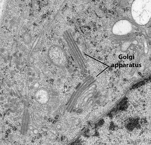
BIOL 230 Lecture Guide - Electron Micrograph of a Golgi Body
Electron Microscope- Definition, Principle, Types, Uses, Labeled Diagram Electron Microscope is in the form of a tall vacuum column that is vertically mounted. It has the following components: 1. Electron gun The electron gun is a heated tungsten filament, which generates electrons. 2. Electromagnetic lenses The condenser lens focuses the electron beam on the specimen.
Solved label the ectron micrograph of an animal cell. | Chegg.com
Muscle Lab 19 Figure 19.5 Sarcomere Diagram | Quizlet THagge TEACHER. Chapter 5 - Tissues Lecture Exam. 61 Terms. THagge TEACHER. Lab 14: Figure 14.10 Anterior Features of the Skull. 9 Terms. THagge TEACHER. Lab 14: Figure 14.11 Lateral View of the Skull. 14 Terms.
Solved and Function PART A ASSESSMENTS Match the terms in ...
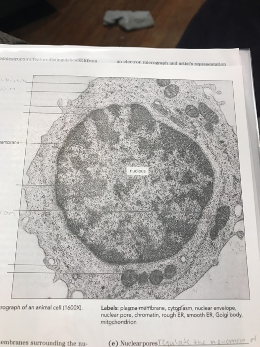
Solved Please label the electron micrograph to assess your | Chegg.com Question: Please label the electron micrograph to assess your knowledge of the structure and function of a cell's nucleus nuclear pore endoplasma reticulum chromatin nucleolus nuclear envelope . This problem has been solved! See the answer See the answer See the answer done loading.

Solved Label the transmission electron micrograph based on ...
Label This Transmission Electron Micrograph : TEM of chloroplast from ... Label the transmission electron micrograph of the nucleus. Provide the labels for the electron micrograph in figure 12.8. Labeling for electron microscopy using antibody conjugated to. Transmission electron microscopy (tem) is a microscopy technique in which a beam of electrons is transmitted through a specimen to form an image.

A scanning electron micrograph of a single granule of ...
Solved: Provide the labels for the electron micrograph in figure 2 ... Answer to Provide the labels for the electron micrograph in figure 2....
![PDF] Evaluation of the Human Enamel Surface Morphology after ...](https://d3i71xaburhd42.cloudfront.net/096d41c63519d122bb265a2a59f00e2c117a44f1/5-Figure2-1.png)
PDF] Evaluation of the Human Enamel Surface Morphology after ...
How to Identify Specimens Embedded in Resin for Electron Microscopy ResiTAG also allows you to track your sections, as they can also be used as labels to identify microscope slides containing the cut sections, with barcodes corresponding to the original capsule or mold. ResiTAG is the most reliable labeling solution for preparing and archiving electron microscopy specimens.
What is a diagram of a plant and animal cell under an ...
Chapter 2 Correlated light and electron microscopy/electron ... - PubMed Electron microscopy and electron tomography provide the highest resolution currently available to study mitochondrial ultrastructure but cannot follow processes in living cells. We describe the combination of these two techniques in which fluorescence confocal microscopy is used to study structural and physiologic changes in mitochondria within ...
Solved Label the transmission electron micrograph based on ...
Concatenated Metallothionein as a Clonable Gold Label for Electron ... The creation of a clonable label could provide the same advantages for electron microscopy provided that the label had high visibility in the electron microscope. Ideally, we want a small protein that would initiate formation of a heavy metal cluster from a heavy metal salt or organometallic compound. We selected the cysteine-rich protein ...
1.2 Ultrastructure of cells assessment
chloroplast and mitochondrion.pdf - D SBA#: Title: Electron micrograph ... At least 2 parts labeled correctly (1 mark) 4 Annotate drawings appropriately and accurately e. 5 parts annotated correctly (4 marks)f. 4 parts annotated correctly (3 marks) g. 3 parts annotated correctly (2 marks)h.

High-magnification electron micrograph of two cross sectioned ...
Electron Micrographs of Cell Organelles - Biology Discussion This is an electron-micrograph of plastid or chloroplast, which is an integral component of all green plant leaves and is characterized by following features (Fig. 15 & 16): (1) They may be spheroidal, ovoid, stellate or collar shaped and differ in size and number in different cells.

Integration of signals from different cortical areas in ...
Scanning electron microscope - Wikipedia A scanning electron microscope (SEM) is a type of electron microscope that produces images of a sample by scanning the surface with a focused beam of electrons.The electrons interact with atoms in the sample, producing various signals that contain information about the surface topography and composition of the sample. The electron beam is scanned in a raster scan pattern, and the position of ...

Electron micrographs of negative staining and immunogold ...
Picking faces out of a crowd: Genetic labels for identification of ... (A) Schematic of indirect (secondary) antibody labeling for correlative light and electron microscopy where the secondary antibody is conjugated to a gold bead or quantum dot. (B) Electron micrograph of microtubules (arrows) labeled with immuno-quantum dots. The primary antibody is an α-tubulin monoclonal antibody.

Muscle Lab 19 Figure 19.5 Sarcomere Diagram | Quizlet
Animal Cell Electron Microscope Labelled - Q14 Draw a large diagram of ... Electron microscopes use accelerated electron beams (as opposed to visible light in a light microscope) to create images of magnification as here is an electron micrograph of an animal cell with the labels superimposed: (i) name the parts labelled as 1 to 10. In the given figure of an animal cell as observed under an electron microscope.
Electron Micrographs
Electron micrographs - plato.acadiau.ca Note: Although this micrograph provides both, only one (size bar or magnification) is usually given. Determining size of a component using a size bar. ... Determine the width of the cell membrane from the transmission electron micrograph provided. Width of membrane = 7-8 nm = 0.0000077 mm ...

A scanning electron micrograph (SEM) of pollen grains from ...
Imaging Specific Protein Labels on Eukaryotic Cells in Liquid with ... (A) Scanning electron microscopy image showing the backside of a microchip with the silicon nitride window in the middle. (B) Schematic of the top view of the slot in the tip of the specimen holder in which the microchips are placed. The microchips are aligned at their sides via alignment poles. The liquid flow path runs through the chips.
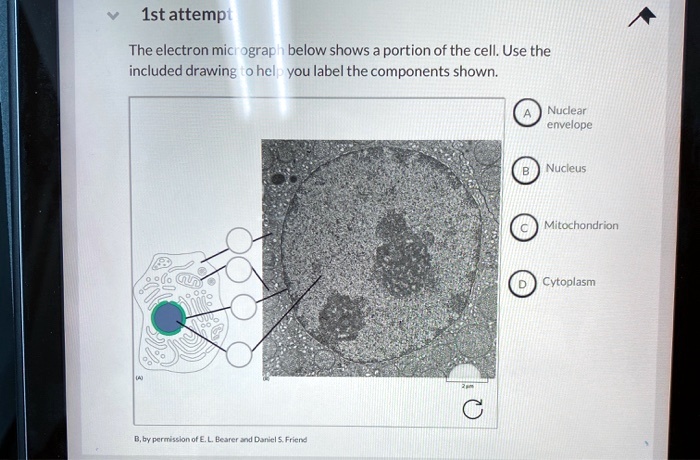
SOLVED:1st attempt The electron micrograph below shows ...
Ultra-Bright and Stable Luminescent Labels for Correlative ... Correlative cathodoluminescence electron microscopy (CCLEM) bioimaging has recently been suggested to provide an attractive alternative based on labels emitting characteristic light. While luminescence excitation by an electron beam enables sub-diffraction imaging, structural damage to the sample by high energy electrons has been identified as ...
Histology Laboratory Manual
Muscle Lab 19 Figure 19.4 Sarcomere Diagram | Quizlet Start studying Muscle Lab 19 Figure 19.4 Sarcomere. Learn vocabulary, terms, and more with flashcards, games, and other study tools.

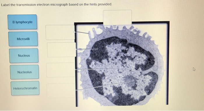
Label electron micrograph of B lymphocyte. - Brainly.com
Label This Transmission Electron Micrograph : TEM of chloroplast … 09-02-2022 · No microtubule labeling is evident. Label this transmission electron micrograph of relaxed sarcomeres by clicking and dragging the labels to the correct location . Molecular labeling for correlative microscopy: And its correlation with the green fluorescent if label (fig. Provide the labels for the electron micrograph in figure 12.8.
WHO Ganti Label Penyebutan Varian COVID-19 - Dari Laut
Plant Cell Nucleus Electron Micrograph : Cell And Organelles Dr Jastrow ... Below is a collection of electron micrographs with labelled subcellular structures that you should be able to identify. In mammals it's average diameter is about 6 an electron micrograph of a section through an animal cell nucleus (from an insect cell). In flowering plants, this condition occurs in sieve tube elements.74.
Electron micrograph showing the distribution of anti-profilin ...
Electron Microscopy SciencesExpiration Labels | Fisher Scientific Manufacturer: Electron Microscopy Sciences 7702405. Quickly identify outdated laboratory items such as reagents and controls. These labels provide space for noting the date that the items were received, opened and when it will expire. A permanent adhesive holds the label firmly in place on a variety of surfaces. The label measures 3/4" x 1 1/2".
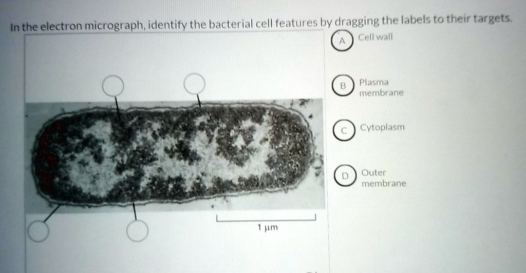
SOLVED:In the electron micrograph; idlentify the bacterial ...
Electron microscope - Wikipedia An electron microscope is a microscope that uses a beam of accelerated electrons as a source of illumination. As the wavelength of an electron can be up to 100,000 times shorter than that of visible light photons, electron microscopes have a higher resolving power than light microscopes and can reveal the structure of smaller objects.. Electron microscopes use shaped magnetic fields to form ...

Electron micrograph showing a neutrophilphagocytosing ...
Electron Micrographs of Cell Organelles | Zoology It is an electron micrograph of cell’s largest and most important organelle – the mitochondria and is characterized by the following features (Fig. 7 & 8): (1) The name mitochondria was given by Benda (1898) and their ma n function was brought to light by Kingsbury (1912). (2) Each mitochondria in section appears as sausage or cup or bowl ...
Prior dengue or yellow fever exposure does not worsen Zika ...

anatomy 10.png - Label the transmission electron micrograph ...
Coloured scanning electron micrograph (SEM) of Plastic wrap ...

Electron Micrograph of Part of a Skeletal Muscle Fiber ...

Transmission electron micrograph (TEM) of norovirus virions ...
Figure 19.1 Srructures found in skeletal muscle fibers (cells ...

Solved Mitochondrion Nucleus Vesicle Peroxisome | Chegg.com

The corresponds to the indicated Learning Out found at the ...

Transmission electron microscope (TEM) micrograph showing the ...
Pre-embedding immunogold labeling to optimize protein ...

Chapter 14 & 15 Flashcards Flashcards | Quizlet
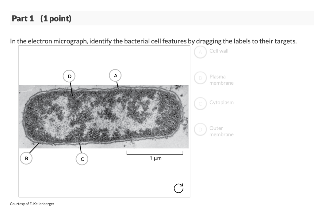
Solved Part 1 (1 point) In the electron micrograph, identify ...
A and B) Electron micrograph of a cell labeled for/5-tubulin ...
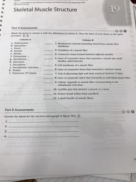
Solved The corresponds to the indicated Learning Out found ...
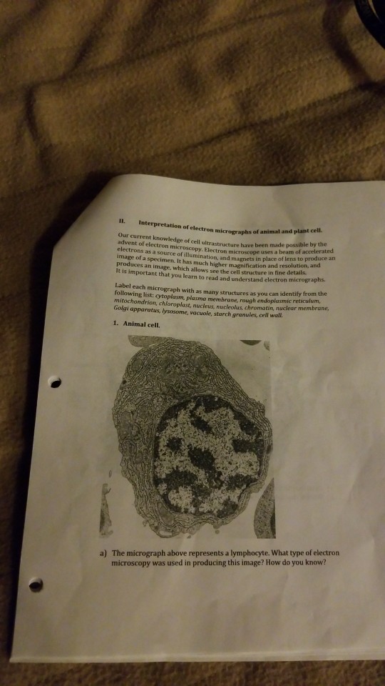
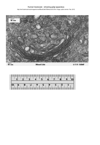
Solved ectron micrographs of animal and plant cell. Our ...
Electron micrograph ultrastructure_magnification

Journal of Helminthology on Twitter: "What is this? A tiny ...

Electron micrograph of an asymmetric synapse from dissociated ...

National Center for Microscopy and Imaging Research | NCMIR
Post a Comment for "42 provide the labels for the electron micrograph"