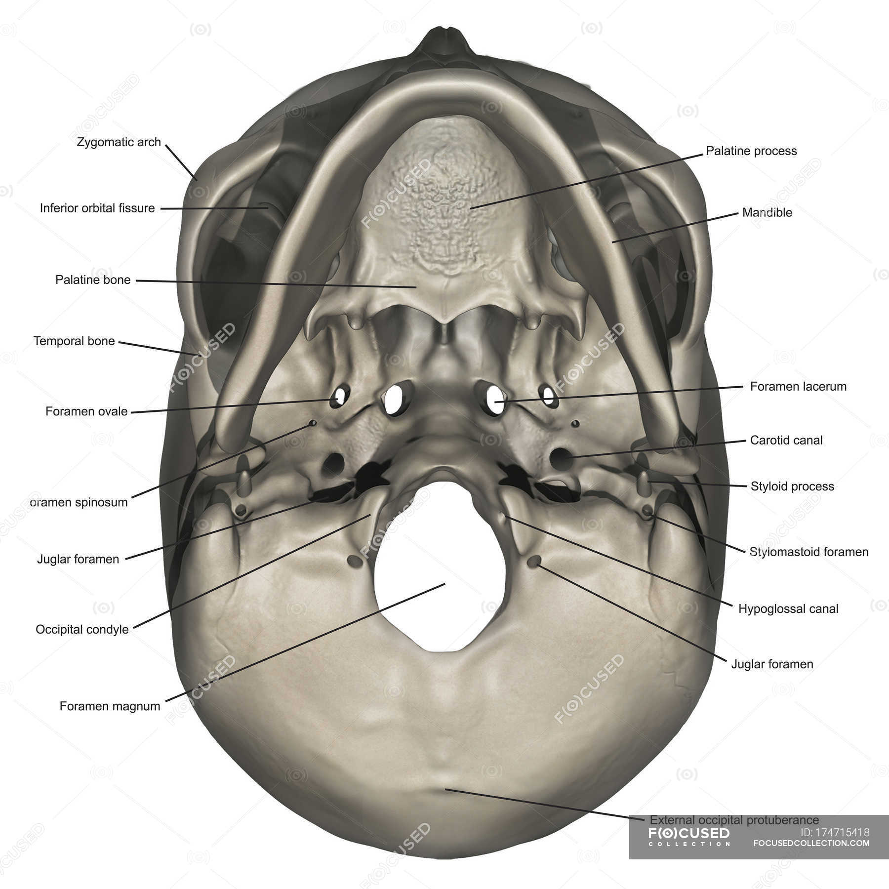
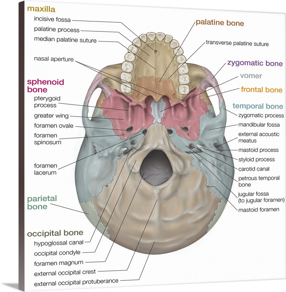
38 inferior skull anatomy labeled
Head and neck anatomy | Radiology Reference Article - Radiopaedia 27.07.2020 · Head and neck anatomy is important when considering pathology affecting the same area. In radiology, the 'head and neck' refers to all the anatomical structures in this region excluding the central nervous system, that is, the brain and spinal cord and their associated vascular structures and encasing membranes i.e. the meninges. Many pathologies are … Carotid canal - Wikipedia Structure. The carotid canal is located within the middle cranial fossa, at the petrous part of the temporal bone.Anteriorly, it is limited by posterior margin of the greater wing of sphenoid bone.Posteromedially, it is limited by basilar part of occipital bone.It is divided in three parts, namely, ascending petrous, transverse petrous, and ascending cavernous parts.
Iliopsoas muscle: Anatomy, function, supply, innervation | Kenhub 19.07.2022 · Contraction of the iliopsoas muscle. The iliopsoas muscle is the strongest flexor of the hip joint. Simultaneous contraction of the psoas major and iliacus muscles produces a powerful flexion of the thigh at the hip joint. However, psoas major can independently act on its attachment on the lumbar spine when its distal end is fixed. Thereby, bilateral contraction of …
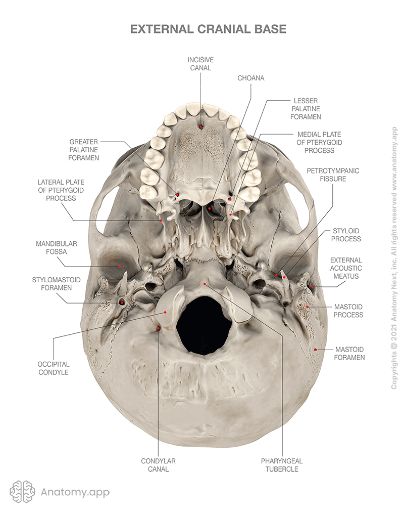
Inferior skull anatomy labeled
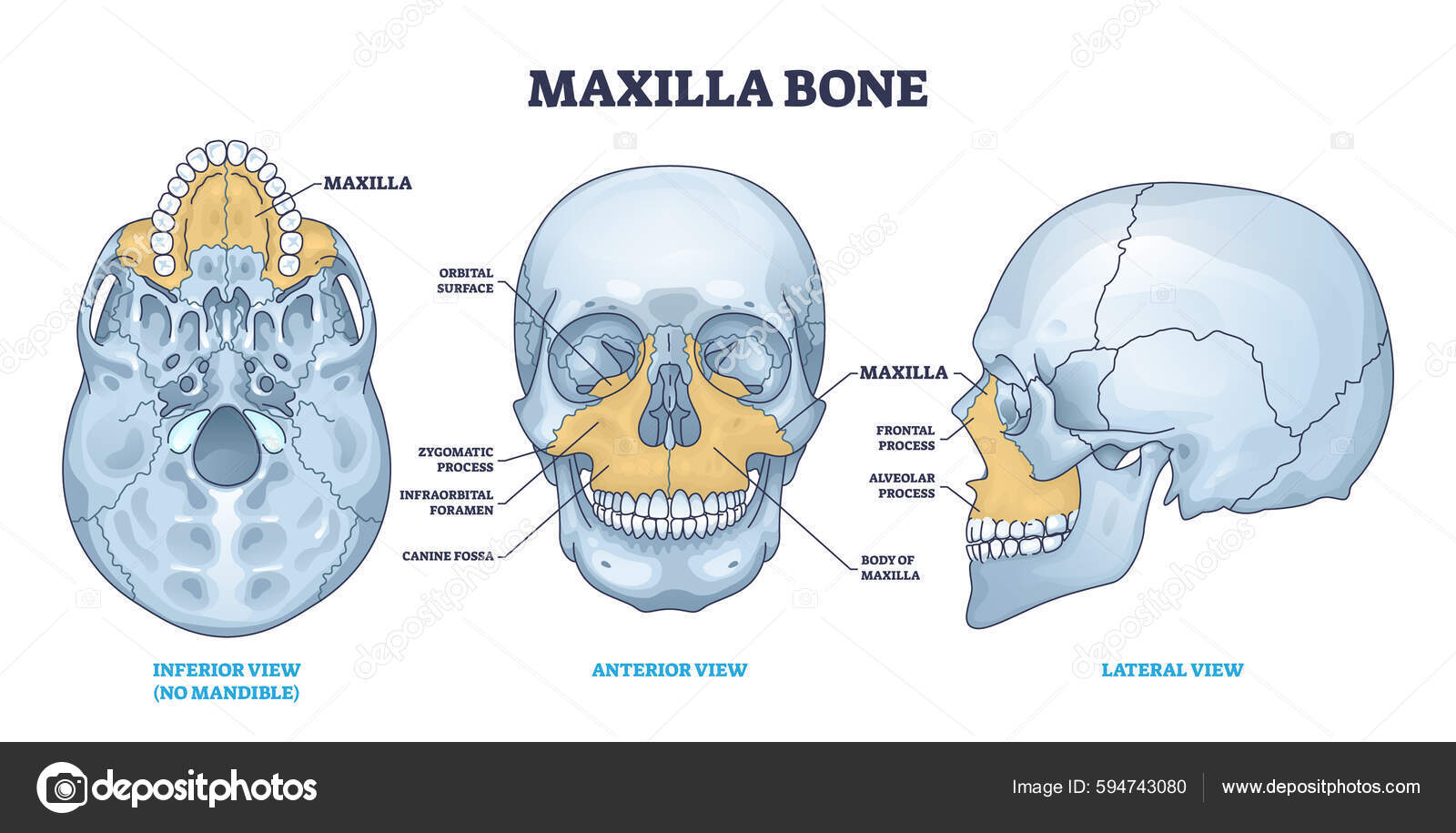
Chapter 7: Integumentary System Flashcards | Quizlet A 32-year-old female is having excision of a mass in her left breast. The physician makes a curved incision along the inferior and medial aspect of the left areola. A breast nodule, measuring approximately 1 cm in diameter, was identified. It appeared to be benign. It was firm, gray, and discrete. It was completely excised. There was no gross ... Palatine process of maxilla - Wikipedia Base of skull. Inferior surface. Roof, floor, and lateral wall of left nasal cavity. Sagittal section of skull. (Palatine process labeled at bottom right.) Medial surface of right maxilla. (Palatine process labeled at center.) External links. Anatomy photo:22:os-1909 at the SUNY Downstate Medical Center – "Osteology of the Skull: The Maxilla" Atlas image: rsa1p7 at the University of Michigan ... Normal chest MDCT with anatomic labels | e-Anatomy - e-Anatomy … 10.03.2022 · Pocket Atlas of Human Anatomy: 5th edition - W. Dauber, Founded by Heinz Fene Anatomical variants and notes from the author about the anatomical labeling of the thorax CT: In the lower lobe of the left lung, there is an inconstant subsuperior pulmonary segment that is seen in approximately 30% of individuals, located between the superior and basal segments of the …
Inferior skull anatomy labeled. Urinary bladder & urethra: Anatomy, location, function | Kenhub 06.07.2022 · The urinary bladder and urethra are pelvic urinary organs whose respective functions are to store and expel urine outside of the body in the act of micturition (urination). As is the case with most of the pelvic viscera, there are differences between male and female anatomy of the urinary bladder and urethra. In our entire urinary system series, the urinary bladder and … Normal chest MDCT with anatomic labels | e-Anatomy - e-Anatomy … 10.03.2022 · Pocket Atlas of Human Anatomy: 5th edition - W. Dauber, Founded by Heinz Fene Anatomical variants and notes from the author about the anatomical labeling of the thorax CT: In the lower lobe of the left lung, there is an inconstant subsuperior pulmonary segment that is seen in approximately 30% of individuals, located between the superior and basal segments of the … Palatine process of maxilla - Wikipedia Base of skull. Inferior surface. Roof, floor, and lateral wall of left nasal cavity. Sagittal section of skull. (Palatine process labeled at bottom right.) Medial surface of right maxilla. (Palatine process labeled at center.) External links. Anatomy photo:22:os-1909 at the SUNY Downstate Medical Center – "Osteology of the Skull: The Maxilla" Atlas image: rsa1p7 at the University of Michigan ... Chapter 7: Integumentary System Flashcards | Quizlet A 32-year-old female is having excision of a mass in her left breast. The physician makes a curved incision along the inferior and medial aspect of the left areola. A breast nodule, measuring approximately 1 cm in diameter, was identified. It appeared to be benign. It was firm, gray, and discrete. It was completely excised. There was no gross ...
Skull | Encyclopedia | Anatomy.app | Learn anatomy | 3D ...

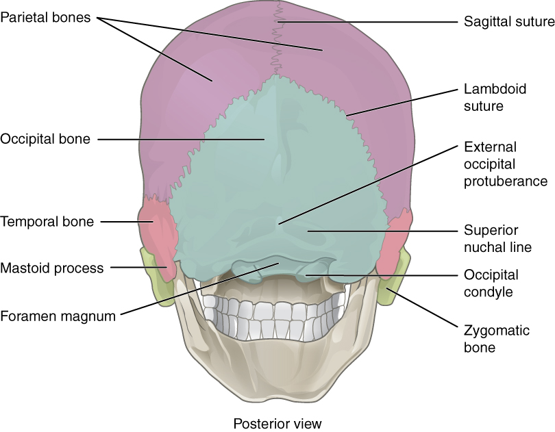
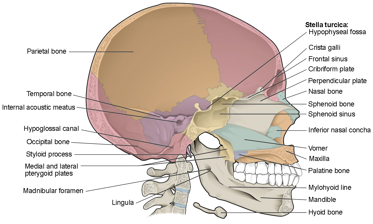
Bones and Features of the Skull – David Fankhauser

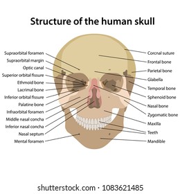
Skull Bones Mnemonic (Cranial and Facial Bones) | Anatomy and Physiology

Skull Anatomy Coloring Sheet

The Skull Bones - Lateral View | GetBodySmart

The Skull Bones Anatomy - Inferior View | GetBodySmart
Inferior view of human skull anatomy with annotations ...

Cranium With Bones Labeled In Anterior And Lateral - Petrous ...
Skull - Knowledge @ AMBOSS
The Skull | Anatomy and Physiology I | | Course Hero
Tinjauan Tulang Mandibula Tanda Tulang Tengkorak Berwarna ...
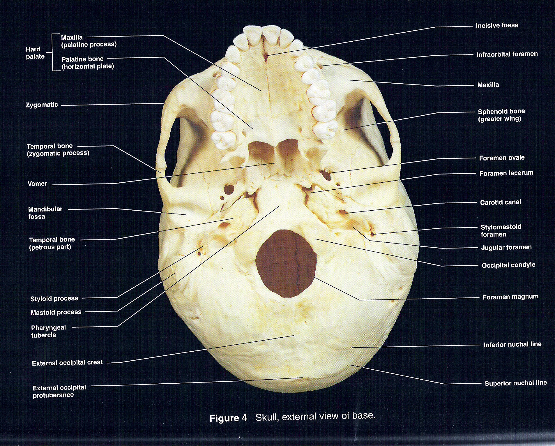
Inferior View of Skull
Skull ○Cranial skeleton (Neurocranium) ○Facial skeleton ...
Vektor Stok Structure Human Skull Main Parts Labeled (Tanpa ...
Learn skull anatomy with skull bone quizzes and diagrams | Kenhub
The Skull | Anatomy and Physiology | | Course Hero
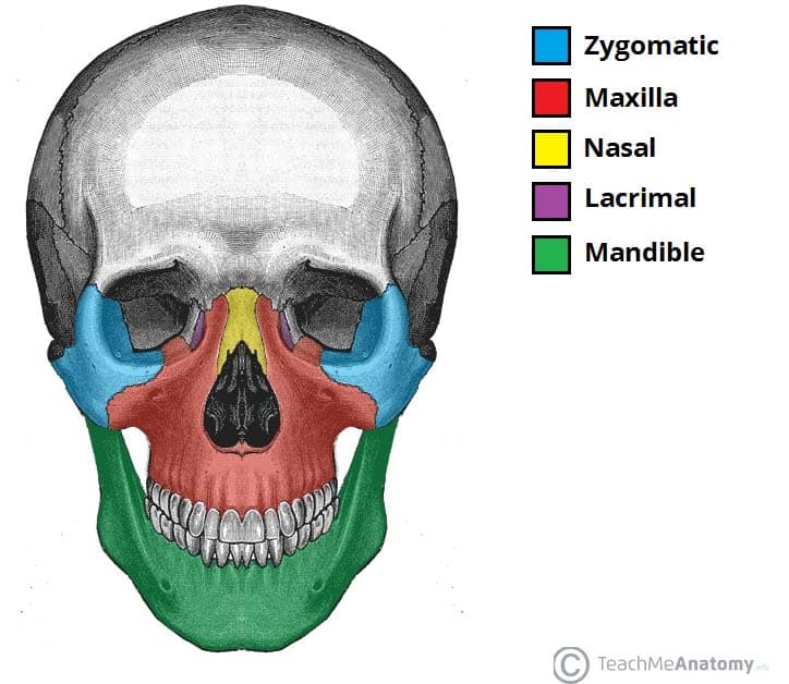
Bones of the Skull - Structure - Fractures - TeachMeAnatomy

Axial CT bone window of skull base from inferior to superior ...

Skull, inferior view | Human anatomy and physiology, Anatomy ...

Multi-colored Skull, inferior view with labels - Axial Ske ...

Skull (Inferior View) - Stock Image - C022/1156 - Science ...
Inferior view of the base of the skull: Anatomy | Kenhub

Solved Please help me label the bones of the skull (inferior ...
The Skull | Anatomy and Physiology I | | Course Hero

Frontal bone | Radiology Reference Article | Radiopaedia.org

Premium Vector | Skull bones head bones skull borders of the ...
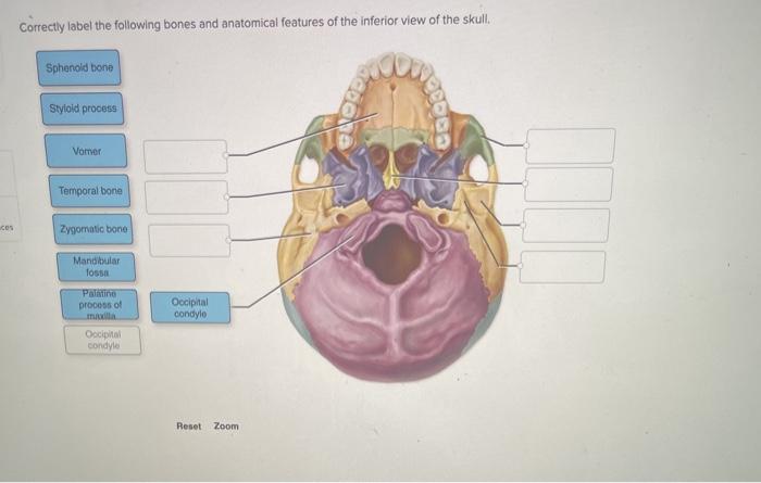
Solved Correctly label the following bones and anatomical ...
Inferior view Vector Art Stock Images | Depositphotos

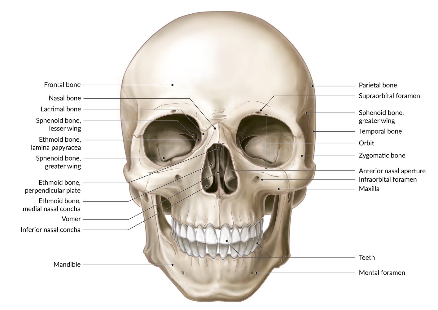
Facial skeleton - Wikipedia
Skull ○Cranial skeleton (Neurocranium) ○Facial skeleton ...

Internal Anatomy of Inferior Skull Part 2 Diagram | Quizlet

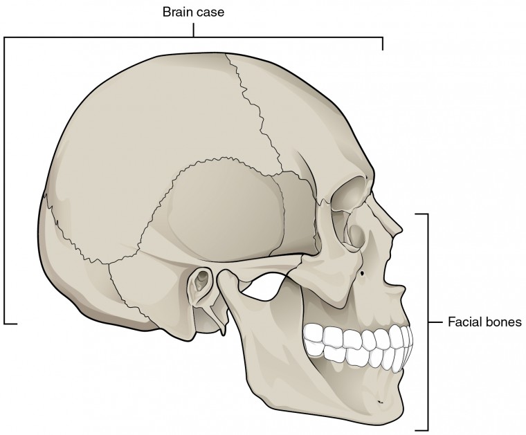
The Skull – Anatomy & Physiology
Inferior view of the base of the skull: Anatomy | Kenhub

Bones and Features of the Skull – David Fankhauser
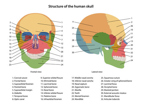
Diagram Of Skull Images – Browse 2,375 Stock Photos, Vectors ...
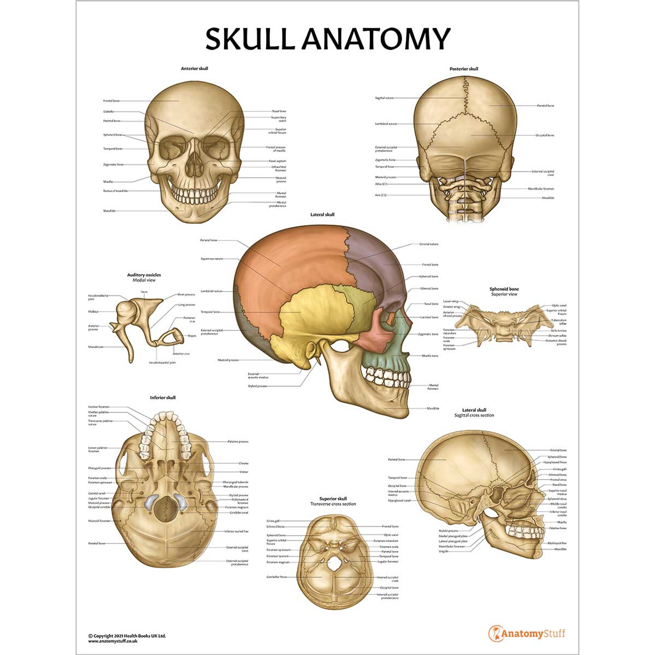
Skull Anatomy Chart / Poster - Laminated
Skull - inferior view. skeletal system Solid-Faced Canvas Print

Anterior Skull Bones Quiz
Post a Comment for "38 inferior skull anatomy labeled"