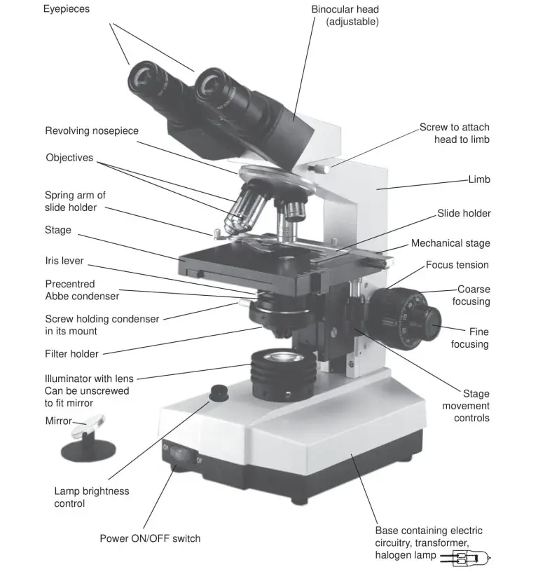
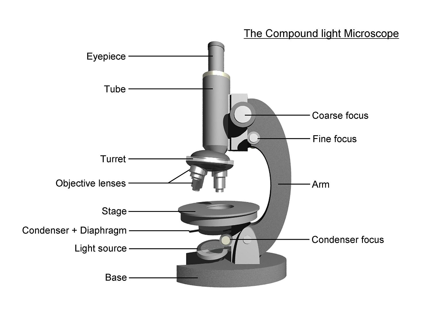
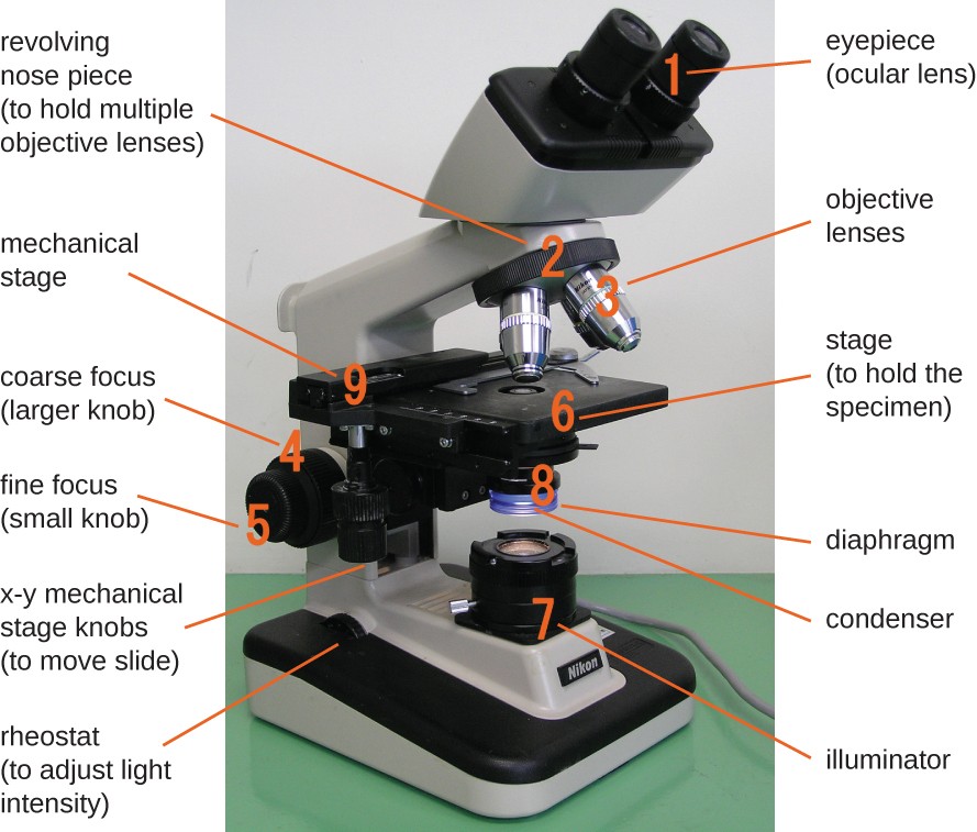
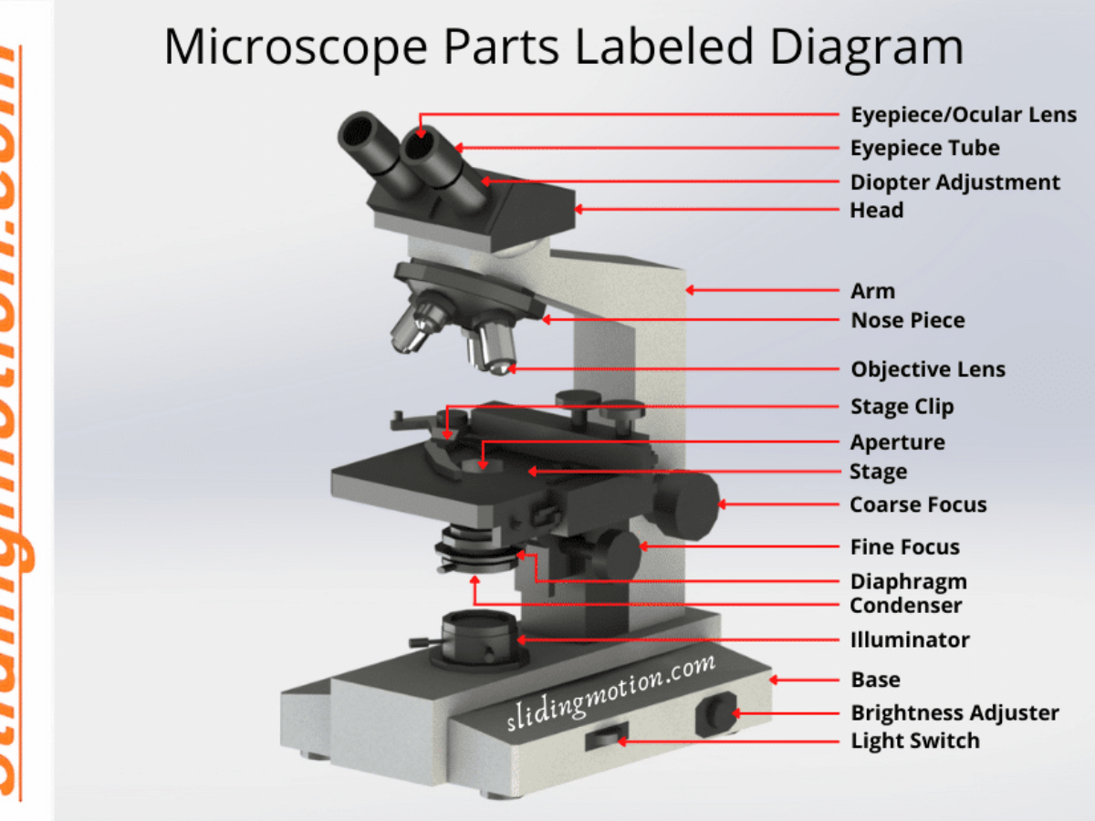
43 diagram of a light microscope with labels
en.wikipedia.org › wiki › Angle_of_viewAngle of view - Wikipedia UV/visible light from an integrating sphere (and/or other source such as a black body) is focused onto a square test target at the focal plane of a collimator (the mirrors in the diagram), such that a virtual image of the test target will be seen infinitely far away by the camera under test. The camera under test senses a real image of the ... en.wikipedia.org › wiki › Thin-film-transistorThin-film-transistor liquid-crystal display - Wikipedia TFT dual-transistor pixel or cell technology is a reflective-display technology for use in very-low-power-consumption applications such as electronic shelf labels (ESL), digital watches, or metering. DTP involves adding a secondary transistor gate in the single TFT cell to maintain the display of a pixel during a period of 1s without loss of ...
› antibody › productDonkey anti-Goat IgG (H+L) Cross-Adsorbed, Alexa Fluor™ 488 ... Asterisks indicate higher magnifications of regions. Combo plots with quantifications of vessel density and numbers of arteries in young and aged pancreas.3D images acquired on a light-sheet microscope of whole cleared young and aged thyroid glands stained with α-SMA and Emcn. Asterisks indicate higher magnifications of regions.

Diagram of a light microscope with labels
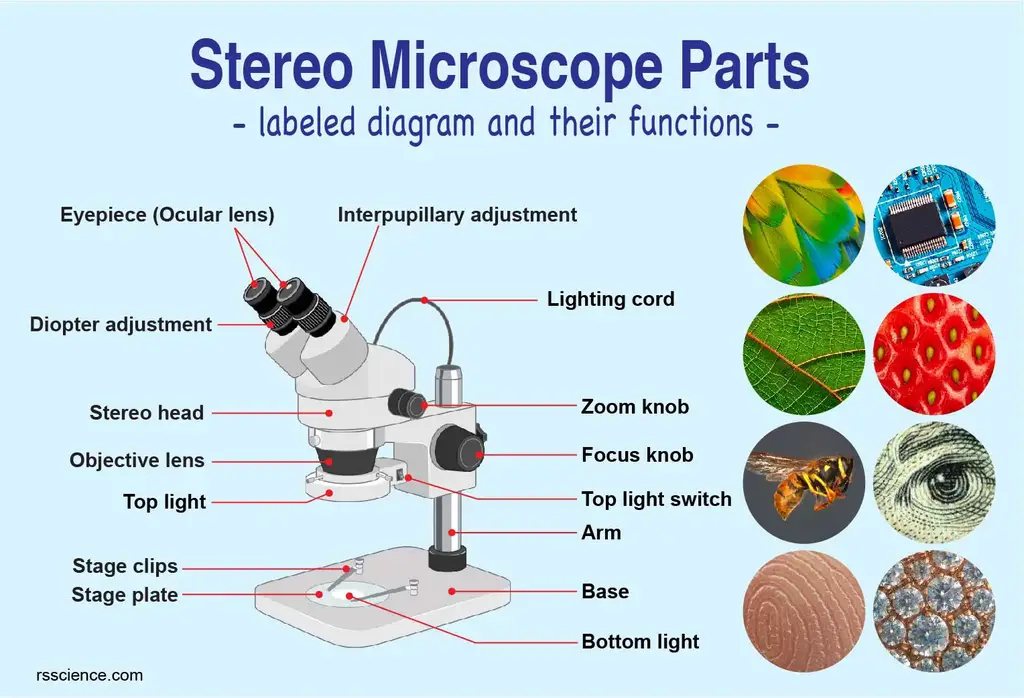
en.wikipedia.org › wiki › Lipid_bilayerLipid bilayer - Wikipedia Lipid bilayers cannot be seen in a traditional microscope because they are too thin. In order to see bilayers, researchers often use fluorescence microscopy. A sample is excited with one wavelength of light and observed in a different wavelength, so that only fluorescent molecules with a matching excitation and emission profile will be seen. › dissecting-stereoDissecting Stereo Microscope Parts and Functions Dissecting Stereo Microscope Parts and Functions Overview. Also known as a stereoscopic microscope, a dissecting microscope is a type of optical microscope commonly used for studying three-dimensional objects (3-D objects) as well as for dissecting biological specimen (e.g. insects and plant parts etc) at low magnification, between 2 and 100x depending on the microscope.
Diagram of a light microscope with labels. › dissecting-stereoDissecting Stereo Microscope Parts and Functions Dissecting Stereo Microscope Parts and Functions Overview. Also known as a stereoscopic microscope, a dissecting microscope is a type of optical microscope commonly used for studying three-dimensional objects (3-D objects) as well as for dissecting biological specimen (e.g. insects and plant parts etc) at low magnification, between 2 and 100x depending on the microscope. en.wikipedia.org › wiki › Lipid_bilayerLipid bilayer - Wikipedia Lipid bilayers cannot be seen in a traditional microscope because they are too thin. In order to see bilayers, researchers often use fluorescence microscopy. A sample is excited with one wavelength of light and observed in a different wavelength, so that only fluorescent molecules with a matching excitation and emission profile will be seen.
Simple Microscope - Parts, Functions, Diagram and Labelling ...
Parts of a Microscope with Their Functions – Microbe Online

Parts of a Microscope - SmartSchool Systems

Simple Microscope Definition, Magnification, Parts And Uses
simple light microscope labeled - Clip Art Library

Compound Microscope Parts – Labeled Diagram and their ...
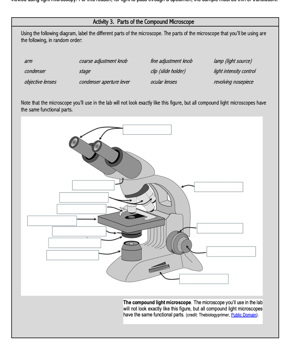
Solved Activity 3. Parts of the Compound Microscope Using ...
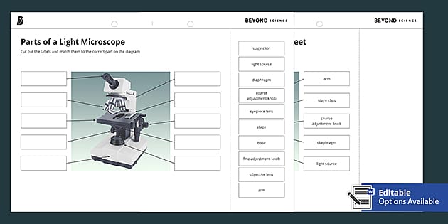
Parts of a Light Microscope Cut and Stick Worksheet - Twinkl
Microscope, Microscope Parts, Labeled Diagram, and Functions

Parts of a Light Microscope Cut and Stick Worksheet - Twinkl

Compound Microscope: Definition, Diagram, Parts, Uses ...
Parts of Stereo Microscope (Dissecting microscope) – labeled ...

Compound Microscope Parts, Functions, and Labeled Diagram ...
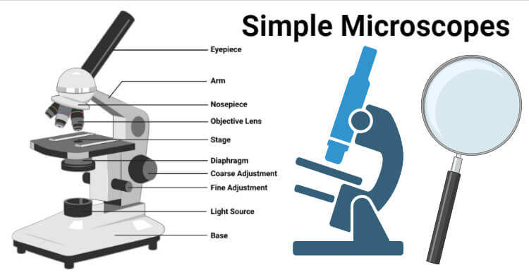
Simple Microscope- Definition, Principle, Magnification ...

Simple Microscope - Diagram (Parts labelled), Principle ...
Instruments of Microscopy | Microbiology | | Course Hero

Compound Microscope Parts – Labeled Diagram and their ...

Compound Microscope- Definition, Labeled Diagram, Principle ...

Parts of a Compound Microscope and Their Functions

Label the microscope — Science Learning Hub

Labelling a Microscope Diagram | Quizlet

Biology Lab Microscope Labeling Diagram | Quizlet

Microscopes: A Beginner's Guide

Microscope With Labels Clip Art at Clker.com - vector clip ...

Microscope With Labels clip art | Microscope parts ...

AP Biology- label standard light microscope Diagram | Quizlet

The Science Break - Labels for the light microscope for GCSE ...

Compound and Stereo- microscopes - Microscopes 4 Schools
16 Basic Parts of Microscope, Function, Names & Labeled Diagram

draw a well label diagram of microscope - Brainly.in

Compound Microscope: Know Definition,working, diagram, properties

Microscope - diagram Tom Butler | Microscope parts, Science ...

Parts of a Microscope Microscope Basics. Label the Compound ...

Compound Microscope Parts – Labeled Diagram and their ...

Microscope diagram labeled | Clipart Panda - Free Clipart Images
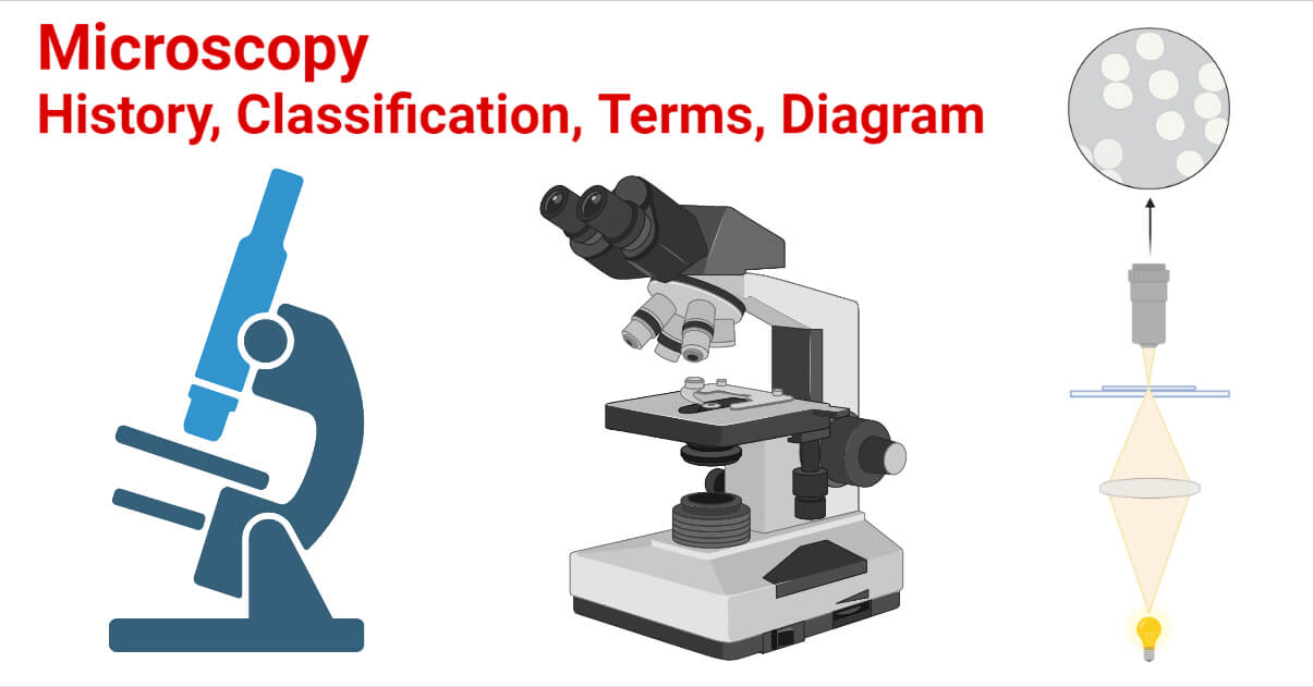
Microscopy- History, Classification, Terms, Diagram
The Compound Light Microscope Label the following parts on ...

Microscope Parts and Functions

Labeling the Parts of the Microscope | Microscope World Resources

Compound Microscope Parts – Labeled Diagram and their ...

Label the light microscope | Teaching Resources
Light Microscope- Definition, Principle, Types, Parts ...

Carl Zeiss Microscopy Optical microscope Worksheet Diagram ...
Post a Comment for "43 diagram of a light microscope with labels"