40 sarcomere labeled
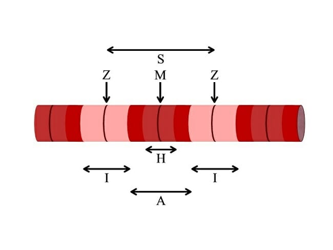
Sarcomeres: “I” and “A” Bands, “M” and “Z” Lines, “H” Zone A sarcomere is a contractile unit of skeletal muscle that is divided into I and A bands, M and Z lines, and H zone. Practice Questions Khan Academy MCAT Official Prep (AAMC) Biology Question Pack, Vol 2. Passage 1 Question 2 Key Points • A sarcomere is the basic contractile unit of skeletal muscle that is made of thick and thin filaments. Sarcomere Labeling Quiz - PurposeGames.com Feb 20, 2023 · Sarcomere Labeling by emcanallen 62,763 plays 8 questions ~20 sec English 8p 24 5.00 (you: not rated) Tries Unlimited [?] Last Played February 20, 2023 - 08:52 PM There is a printable worksheet available for download here so you can take the quiz with pen and paper. Remaining 0 Correct 0 Wrong 0 Press play! 0% 10:00.0 Show More
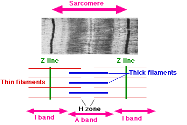
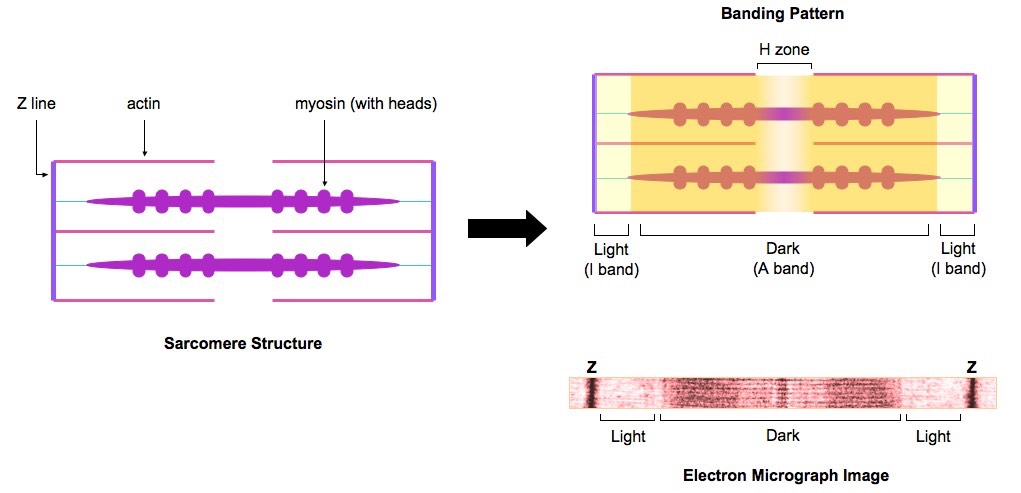
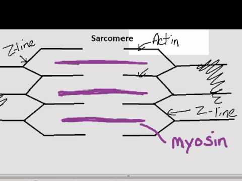
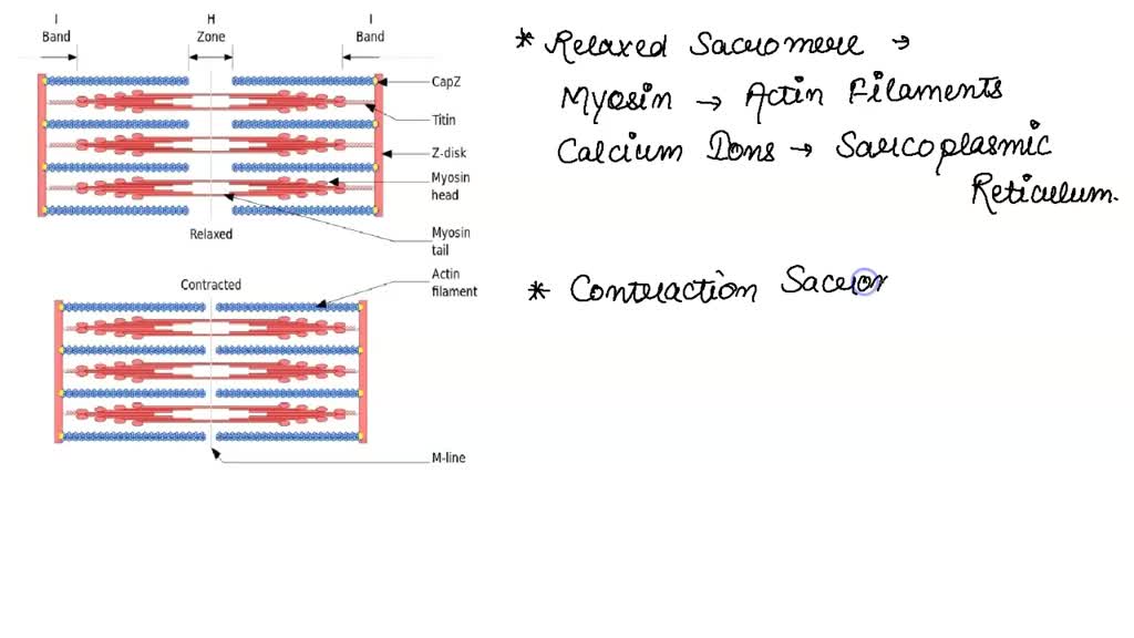
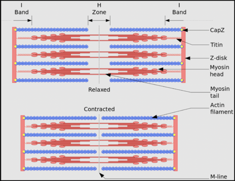
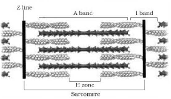
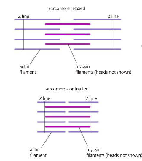
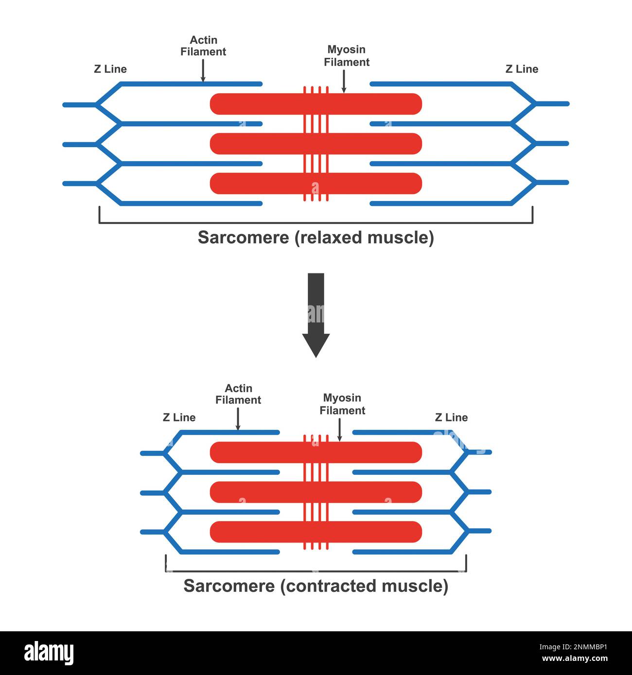
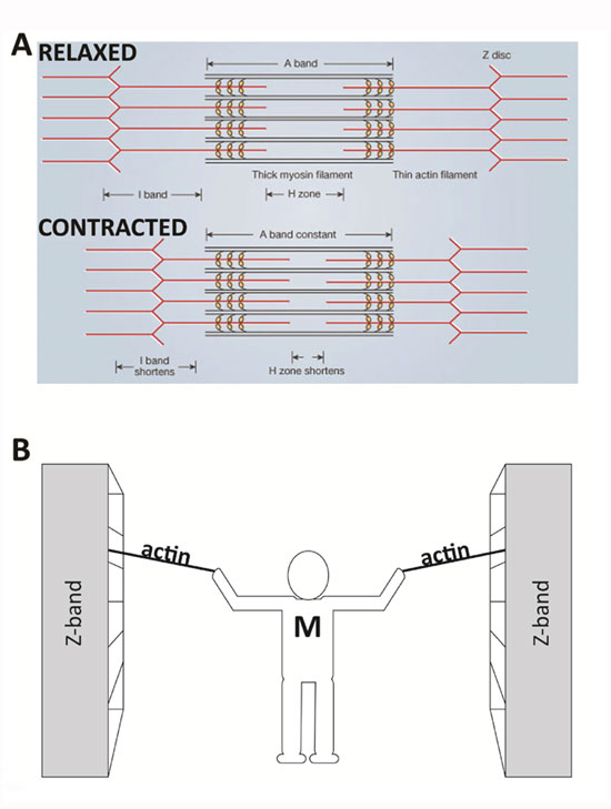
Sliding Filament Model of Contraction | Biology for Majors II A sarcomere is defined as the distance between two consecutive Z discs or Z lines; when a muscle contracts, the distance between the Z discs is reduced. The H zone—the central region of the A zone—contains only thick filaments and is shortened during contraction. The I band contains only thin filaments and also shortens.
Sarcomere labeled
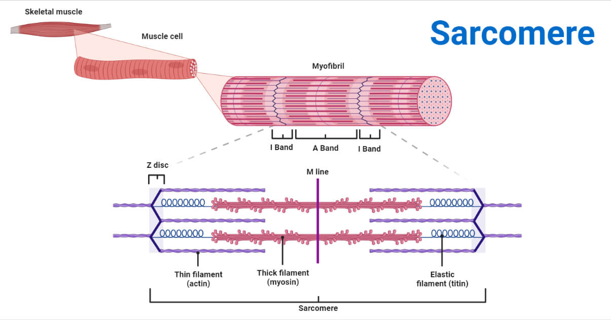
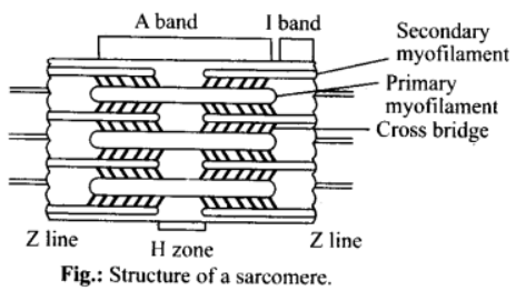
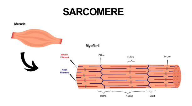
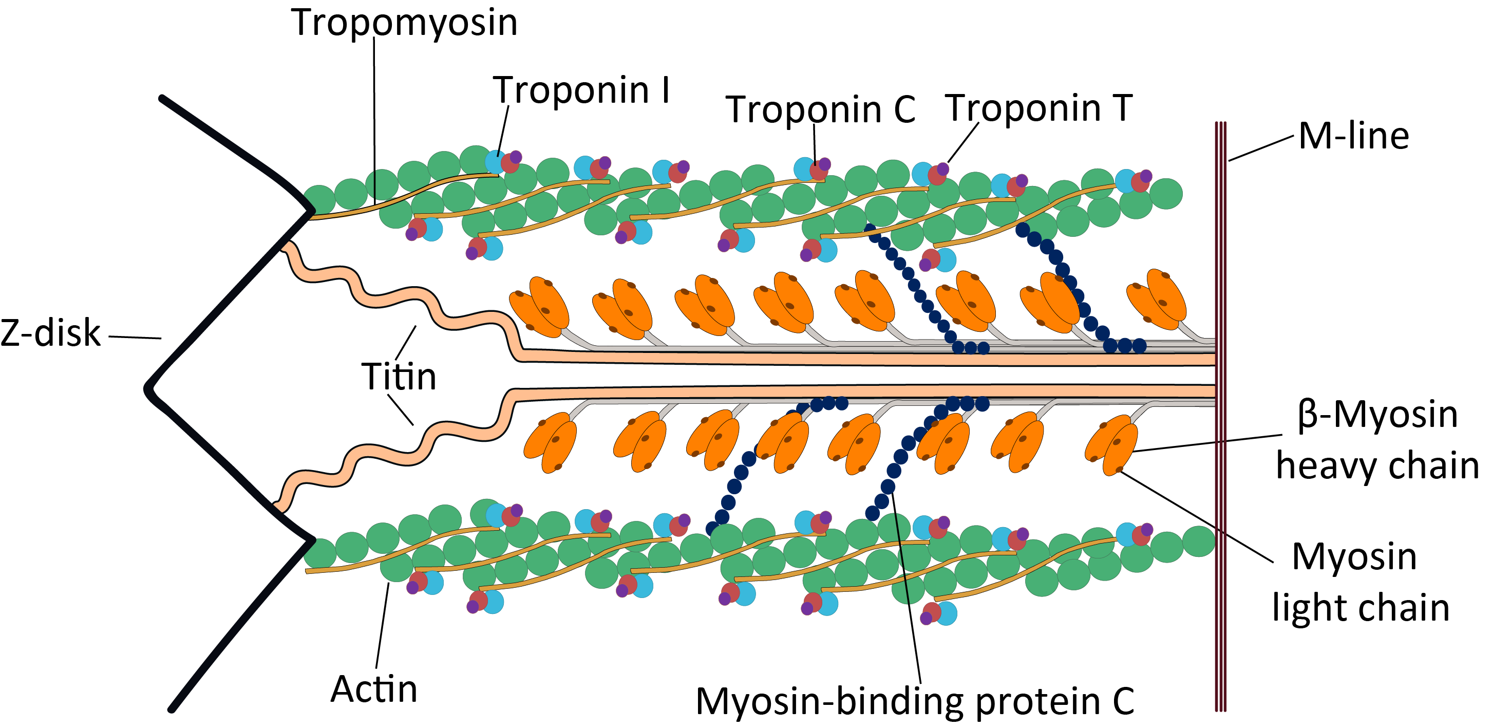
9.1C: Sliding Filament Model of Contraction - Medicine LibreTexts A sarcomere is defined as the segment between two neighboring, parallel Z-lines. Z lines are composed of a mixture of actin myofilaments and molecules of the highly elastic protein titin crosslinked by alpha-actinin. Actin myofilaments attach directly to the Z-lines, whereas myosin myofilaments attach via titin molecules. What is a Sarcomere? - Parts & Contraction - Study.com A sarcomere is a basic unit of function within muscle tissue. Muscle sarcomeres are arranged in stacked patterns throughout each muscle. They help to coordinate muscle movements, especially ... 10.2 Skeletal Muscle - Anatomy & Physiology A sarcomere is defined as the region of a myofibril contained between two cytoskeletal structures called Z-discs (also called Z-lines or Z-bands), and the striated appearance of skeletal muscle fibers is due to the arrangement of the thick and thin myofilaments within each sarcomere ( Figure 10.2.2 ).
Sarcomere labeled. Labeled Sarcomere Diagram A sarcomere is the basic unit of striated muscle tissue. It is the repeating unit between two Z lines. Skeletal muscles are composed of tubular muscle cells which. Sarcomeres are composed of thick filaments and thin filaments. The thin filaments Look at the diagram above and realize what happens as a muscle contracts. Sarcomere - an overview | ScienceDirect Topics A sarcomere is the functional unit (contractile unit) of a muscle fiber. As illustrated in Figure 2-5, each sarcomere contains two types of myofilaments: thick filaments, composed primarily of the contractile protein myosin, and thin filaments, composed primarily of the contractile protein actin. Sarcomere Labeling Diagram | Quizlet Sarcomere Definition The smallest contractile unit of muscle; extends from one Z disc to the next Location Term H Band Definition The band at the middle of the A Band, where only myosin is found Location Term A Band Definition The darkest area that runs the length of the myosin, including where actin and myosin overlap Location Term I Band Sarcomere Definition & Meaning - Merriam-Webster sarcomere noun sar· co· mere ˈsär-kə-ˌmi (ə)r : any of the repeating, contractile, structural subunits of striated muscle cells (as of skeletal or cardiac muscle) that are composed of the protein filaments actin and myosin Skeletal muscle is a unique kind of tissue, made up of long, thin fibers composed of several different proteins.
Comparison of a relaxed and contracted sarcomere Comparison of a relaxed and contracted sarcomere (A) The basic organization of a sarcomere subregion, showing the centralized location of myosin (A band). Actin and the z discs are shown in... Sarcomere - Wikipedia A sarcomere (Greek σάρξ sarx "flesh", μέρος meros "part") is the smallest functional unit of striated muscle tissue. [1] It is the repeating unit between two Z-lines. Skeletal muscles are composed of tubular muscle cells (called muscle fibers or myofibers) which are formed during embryonic myogenesis. Myofibril - Definition, Function and Structure | Biology Dictionary Myofibril Definition. A myofibril is a component of the animal skeletal muscle. Myofibrils are long filaments that run parallel to each other to form muscle (myo) fibers. The myofibrils, and resulting myofibers, may be several centimeters in length. The muscle fibers are single multinucleated cells that combine to form the muscle. Sarcomere Model Sarcomere Structure - YouTube This video was produced to help students of human anatomy at Modesto Junior College study our anatomical models.
Sarcomere- Definition, Structure, Diagram, and Functions Jul 7, 2022 · A sarcomere is a complex multicomponent biological system and functional unit of striated muscle which plays a vital role in transforming the chemical energy released upon the ATP hydrolysis into mechanical work. Skeletal muscles are made up of the basic unit called a sarcomere and all voluntary movement is initiated by this skeletal muscle. What Is The Sliding Filament Theory? - BYJU'S The sliding filament theory is a suggested mechanism of contraction of striated muscles, actin and myosin filaments to be precise, which overlap each other resulting in the shortening of the muscle fibre length. Actin (thin) filaments combined with myosin (thick filaments) conduct cellular movements. Myosin is a protein that converts ATP ... Sarcomere - Definition, Structure, Function and Quiz - Biology Dictionary A sarcomere is the functional unit of striated muscle. This means it is the most basic unit that makes up our skeletal muscle. Skeletal muscle is the muscle type that initiates all of our voluntary movement. Herein lies the sarcomere's main purpose. Sarcomeres are able to initiate large, sweeping movement by contracting in unison. The Sarcomere and Sliding Filaments in Muscular Contraction: Definition ... A given myofibril contains approximately 10,000 sarcomeres, each of which is about 3 micrometers in length. While each sarcomere is small, several sarcomeres added together span the length of the ...
Sarcomere - Physiopedia
Anatomy of a skeletal muscle fiber (video) | Khan Academy When the muscle fiber is relaxed , the contact surface between both types of protein filaments is at a minumum, When the fiber is recieves a nerve stimulus, the thin. filaments slide over the thick filaments, causing distance between the Z lines, Which makes up the boundary of the sarcomere, to become narrow.

Sarcomere - an overview | ScienceDirect Topics
Sarcomere Structure : Mnemonic | Epomedicine Schematic representation of a sarcomere. Z is the final alphabet: Z lines represents the end of sarcomere. M for middle: M line represents the midline of sarcomere. I is a thin letter: I band has only thin filaments. H is a thick letter: H zone has only thick filaments. A is a hybrid of "I" and "H": A band has both thin and thick ...
Sarcomeres | BioNinja
Skeletal muscle tissue: Histology | Kenhub The sarcomere is the functional unit of a skeletal muscle cell. Each sarcomere is about 2.5 micrometers in length. It is made up of multiple myosin and actin filaments oriented in parallel. The actin and myosin filaments overlap in certain places creating several bands and zones. A Z disc forms the boundary of the sarcomere on either side. Thin ...

4 Schematic diagram of a single sarcomere: (above) in ...
Sliding Filament Theory, Sarcomere, Muscle Contraction, Myosin |... Sarcomeres are highly stereotyped and are repeated throughout muscle cells, and the proteins within them can change in length, which causes the overall length of a muscle to change. An individual...
Parts of the Sarcomere
Sarcomere - Physiopedia A sarcomere is the basic contractile unit of a myocyte (muscle fibre). A sarcomere is composed of two main protein filaments (thin actin and thick myosin filaments) which are the active structures responsible for muscular contraction. The widely accepted theory describing muscular contraction is called the sliding filament theory, which proposes that the active force is generated as actin ...

Sarcomeres — Science Learning Hub
Sarcomere | Definition, Structure, & Sliding Filament Theory Aug 10, 2019 · A sarcomere describes as the distance between two Z discs or Z lines. When a muscle contracts in our body the distance reduces between the Z discs. The central region of the A zone (H zone), contains only thick filaments (myosin), and became short during contraction.

302 Sarcomere Images, Stock Photos & Vectors | Shutterstock
Sarcomere: anatomy, structure and function | Kenhub The sarcomere is the main contractile unit of muscle fiber in the skeletal muscle. Each sarcomere is composed of protein filaments ( myofilaments) that include mainly the thick filaments called myosin, and thin filaments called actin. The bundles of myofilaments are called myofibrils .

Diagram of sarcomere - Brainly.in
Label the Sarcomere Structure Diagram | Quizlet Label the Sarcomere Structure Learn Test Match Created by jack_burton76 Terms in this set (12) Term z disc Location Term mysosin (thick) Location Term thin (actin) filament Location Term I band Location Term A band Location Term I band Location Term H zone Location Term elastic (titin) filaments Location Term elastic (titin) filaments Location Term

Sarcomere - an overview | ScienceDirect Topics
Sarcomeres | BioNinja Myofibrils consist of repeating contractile units called sarcomeres, which are made of two protein myofilaments The thick filament (myosin) contains small protruding heads which bind to regions of the thin filament (actin) Movement of these two filaments relative to one another causes the lengthening and shortening of the sarcomere

Sarcomere Labeling Quiz
210+ Sarcomere Stock Photos, Pictures & Royalty-Free Images - iStock Sarcomere muscular biology scheme vector illustration Sarcomere muscular biology scheme vector illustration. Myosin filaments, discs, lines and bands. Myofibril detailed labeled diagram. Sports educational health information. Muscular system anatomy. sarcomere stock illustrations

Muscle Structure And Control Of Contraction - Muscle System ...
Sarcomere Illustrations & Vectors - Dreamstime Download 77 Sarcomere Stock Illustrations, Vectors & Clipart for FREE or amazingly low rates! New users enjoy 60% OFF. 207,311,803 stock photos online. ... Labeled skeletal muscle anatomy vector illustration drawing. Free with trial. Histology of human skeletal muscle under microscope view.
Sarcomere- Definition, Structure, Diagram, and Functions
sarcomere labeled diagram Diagram | Quizlet sarcomere labeled diagram Flashcards Learn Test Match Created by ruthiewilhelm Terms in this set (8) Term i band Definition light band Location Term m line Definition middle of sarcomere Location Term myosin Definition thick filament Location Term a band Definition dark band Location Term h zone Definition only myosin Location Term actin Definition
How Muscle Fibers stretch? What happens on the level of ...
10.2 Skeletal Muscle - Anatomy & Physiology A sarcomere is defined as the region of a myofibril contained between two cytoskeletal structures called Z-discs (also called Z-lines or Z-bands), and the striated appearance of skeletal muscle fibers is due to the arrangement of the thick and thin myofilaments within each sarcomere ( Figure 10.2.2 ).

Schematic diagram of a muscle sarcomere. The isotropic and ...
What is a Sarcomere? - Parts & Contraction - Study.com A sarcomere is a basic unit of function within muscle tissue. Muscle sarcomeres are arranged in stacked patterns throughout each muscle. They help to coordinate muscle movements, especially ...
Draw and label a diagram of a sarcomere in a relaxed myofibril., Include the positions of the thick and thin filaments and the, orientation of the cytoskeletal components. Below this, draw a, similar ...
9.1C: Sliding Filament Model of Contraction - Medicine LibreTexts A sarcomere is defined as the segment between two neighboring, parallel Z-lines. Z lines are composed of a mixture of actin myofilaments and molecules of the highly elastic protein titin crosslinked by alpha-actinin. Actin myofilaments attach directly to the Z-lines, whereas myosin myofilaments attach via titin molecules.
Sarcomere - Physiopedia
Draw the diagram of a sarcomere of skeletal muscle showing ...
SKELETAL MUSCLE ORGANIZATION
Draw the diagram of a sarcomere of skeletal muscle showing ...
11.2: Movement - The!Mad!Scientist!

Sliding filament theory - Wikipedia

302 Sarcomere Images, Stock Photos & Vectors | Shutterstock

Sarcomeres | BioNinja
Sarcomere hi-res stock photography and images - Alamy

Schematic representation of a sarcomere. The thick and thin ...

Sarcomere Labeled | Science diagrams, Labels, Student learning
Sliding Filament Theory, Sarcomere, Muscle Contraction ...

Lapisan Struktur Otot Rangka Dengan Closeup Anatomi ...

Draw the diagram of a sarcomere of skeletal muscle showing different regions | 11 | LOCOMOTION A...
Solved] Draw a diagram of the components of a sarcomere ...
![2. The [sarcomere] length-tension relation](http://www.bristol.ac.uk/phys-pharm-neuro/media/plangton/ugteach/ugindex/m1_index/nm_tension/image/lt_long.gif)
2. The [sarcomere] length-tension relation

Complete Giant Sarcomere Model by Denoyer-Geppert - Anatomy ...
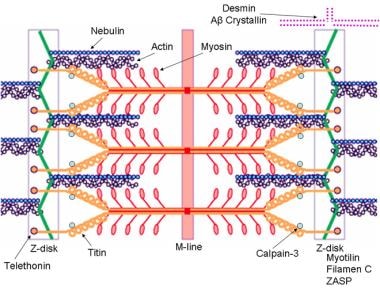
Muscular Dystrophy: Practice Essentials, Pathophysiology ...

Muscle tissue - Knowledge @ AMBOSS
Sarcomere - Definition, Structure, Function and Quiz ...

ImageQuiz: Muscle sarcomere structure
50+ Sarcomere Illustrations, Royalty-Free Vector Graphics ...
File:Cardiac sarcomere structure.png - Wikipedia
Image: Sarcomere

Structure Sarcomere Vector & Photo (Free Trial) | Bigstock
Post a Comment for "40 sarcomere labeled"